Explore
 Validate
 Learn
Learn Western blot
Antibody data
- Antibody Data
- Antigen structure
- References [1]
- Comments [0]
- Validations
- Western blot [4]
- Immunocytochemistry [2]
- Immunoprecipitation [1]
- Immunohistochemistry [3]
Submit
Validation data
Reference
Comment
Report error
- Product number
- GTX106012 - Provider product page

- Provider
- GeneTex
- Proper citation
- GeneTex Cat#GTX106012, RRID:AB_11176384
- Product name
- Rad21 antibody
- Antibody type
- Polyclonal
- Reactivity
- Human, Mouse
- Host
- Rabbit
Submitted references RAD50 phosphorylation promotes ATR downstream signaling and DNA restart following replication stress.
Gatei M, Kijas AW, Biard D, Dörk T, Lavin MF
Human molecular genetics 2014 Aug 15;23(16):4232-48
Human molecular genetics 2014 Aug 15;23(16):4232-48
No comments: Submit comment
Supportive validation
- Submitted by
- GeneTex (provider)
- Main image
- Experimental details
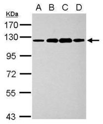
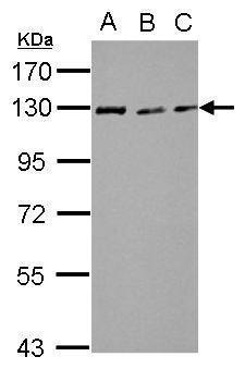
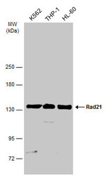
- Sample (30 ug of whole cell lysate) A: K562 B: THP-1 C: HL-60 D: NCI-H929 7.5% SDS PAGE GTX106012 diluted at 1:1000
- Validation comment
- WB
- Submitted by
- GeneTex (provider)
- Main image
- Experimental details
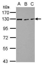
- Sample (30 ?g of whole cell lysate) A: NIH-3T3 B: JC C: BCL-1 7.5% SDS PAGE GTX106012 diluted at 1:3000 The HRP-conjugated anti-rabbit IgG antibody (GTX213110-01) was used to detect the primary antibody.
- Submitted by
- GeneTex (provider)
- Main image
- Experimental details
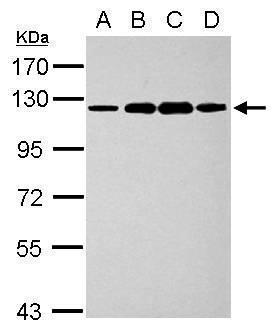
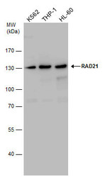
- RAD21 antibody detects RAD21 protein by western blot analysis. Various whole cell extracts (30 ?g) were separated by 7.5% SDS-PAGE, and the membrane was blotted with RAD21 antibody (GTX106012) diluted by 1:1000. The HRP-conjugated anti-rabbit IgG antibody (GTX213110-01) was used to detect the primary antibody.
- Submitted by
- GeneTex (provider)
- Main image
- Experimental details
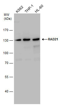
- Various whole cell extracts (30 ?g) were separated by 5% SDS-PAGE, and the membrane was blotted with Rad21 antibody (GTX106012) diluted at 1:1000. The HRP-conjugated anti-rabbit IgG antibody (GTX213110-01) was used to detect the primary antibody.

Supportive validation
- Submitted by
- GeneTex (provider)
- Main image
- Experimental details
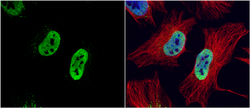
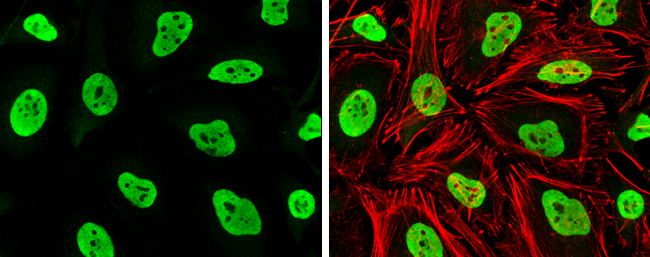
- RAD21 antibody detects RAD21 protein at nucleus by immunofluorescent analysis.Sample: HeLa cells were fixed in 4% paraformaldehyde at RT for 15 min.Green: RAD21 protein stained by RAD21 antibody (GTX106012) diluted at 1:1000.Red: alpha Tubulin, a cytoskeleton marker, stained by alpha Tubulin antibody [B-5-1-2] (GTX11304) diluted at 1:10000.Blue: Hoechst 33342 staining.
- Submitted by
- GeneTex (provider)
- Main image

- Experimental details
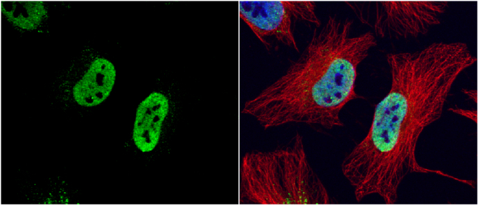
- Rad21 antibody detects Rad21 protein at nucleus by immunofluorescent analysis.Sample: HeLa cells were fixed in 4% paraformaldehyde at RT for 15 min.Green: Rad21 stained by Rad21 antibody (GTX106012) diluted at 1:1000.Red: phalloidin, a cytoskeleton marker, diluted at 1:100.
Supportive validation
- Submitted by
- GeneTex (provider)
- Main image
- Experimental details
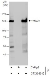
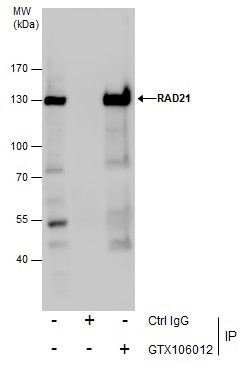
- Immunoprecipitation of RAD21 protein from Jurkat whole cell extracts using 5 £gg of RAD21 antibody (GTX106012).Western blot analysis was performed using RAD21 antibody (GTX106012).EasyBlot anti-Rabbit IgG (GTX221666-01) was used as a secondary reagent.
Supportive validation
- Submitted by
- GeneTex (provider)
- Main image
- Experimental details
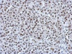
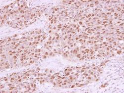
- Immunohistochemical analysis of paraffin-embedded HBL435 xenograft, using RAD21(GTX106012) antibody at 1:750 dilution.
- Submitted by
- GeneTex (provider)
- Main image
- Experimental details
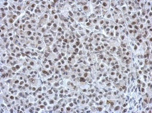
- Immunohistochemical analysis of paraffin-embedded C2C12 xenograft, using RAD21(GTX106012) antibody at 1:750 dilution.
- Submitted by
- GeneTex (provider)
- Main image
- Experimental details
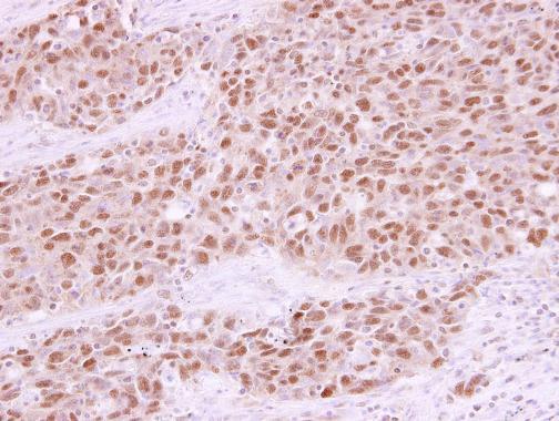
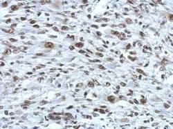
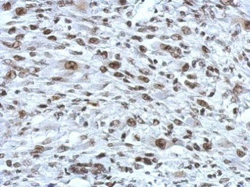
- RAD21 antibody detects RAD21 protein at nucleus in human lung papillary adenocarcinoma by immunohistochemical analysis. Sample: Paraffin-embedded human lung papillary adenocarcinoma. RAD21 antibody (GTX106012) diluted at 1:250.