 Explore
 Validate
 Learn
LearnSTJ11101535
antibody from St John's Laboratory
Targeting: RAD21
hHR21, KIAA0078, SCC1
 Western blot
 Immunocytochemistry Immunoprecipitation Immunohistochemistry Chromatin Immunoprecipitation
Antibody data
- Antibody Data
- Antigen structure
- References [0]
- Comments [0]
- Validations
- Western blot [2]
- Immunohistochemistry [4]
- Chromatin Immunoprecipitation [2]
Submit
Validation data
Reference
Comment
Report error
- Product number
- STJ11101535 - Provider product page

- Provider
- St John's Laboratory
- Product name
- Anti-RAD21 antibody (STJ11101535)
- Antibody type
- Polyclonal
- Description
- Rabbit polyclonal antibody anti-RAD21 is suitable for use in Western Blot, Immunohistochemistry, Immunofluorescence and Immunoprecipitation.
- Reactivity
- Human, Mouse, Rat
- Host
- Rabbit
- Conjugate
- Unconjugated
- Antigen sequence
NA- Epitope
- NA
- Isotype
- IgG
- Antibody clone number
- NA
- Vial size
- NA
- Concentration
- NA
- Storage
- Store in a freezer at-20°C and avoid freeze-thaw cycles.
- Handling
- NA
No comments: Submit comment
Supportive validation
Supportive validation
- Submitted by
- St John's Laboratory (provider)
- Main image

- Experimental details
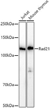
- Western blot analysis of extracts of various cell lines, using Rad21 antibody (STJ11101535) at 1:1000 dilution. Secondary antibody: HRP Goat Anti-rabbit IgG (H+L) at 1:10000 dilution. Lysates/proteins: 25ug per lane. Blocking buffer: 3% nonfat dry milk in TBST. Detection: ECL Basic Kit. Exposure time: 10s.
- Sample type
- NA
- Validation comment
- NA
- Primary Ab dilution
- NA
- Other comments
- NA
- Secondary Ab
- NA
- Secondary Ab dilution
- NA
- Protocol
- NA
Supportive validation
- Submitted by
- St John's Laboratory (provider)
- Main image

- Experimental details
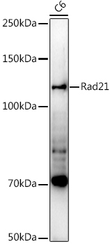
- Western blot analysis of extracts of C6 cells, using Rad21 antibody (STJ11101535) at 1:1000 dilution. Secondary antibody: HRP Goat Anti-rabbit IgG (H+L) at 1:10000 dilution. Lysates/proteins: 25ug per lane. Blocking buffer: 3% nonfat dry milk in TBST. Detection: ECL Enhanced Kit. Exposure time: 90s.
- Sample type
- NA
- Validation comment
- NA
- Primary Ab dilution
- NA
- Other comments
- NA
- Secondary Ab
- NA
- Secondary Ab dilution
- NA
- Protocol
- NA
Supportive validation
Supportive validation
Supportive validation
Supportive validation
- Submitted by
- St John's Laboratory (provider)
- Main image
- Experimental details
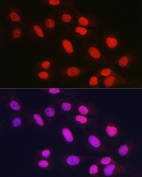
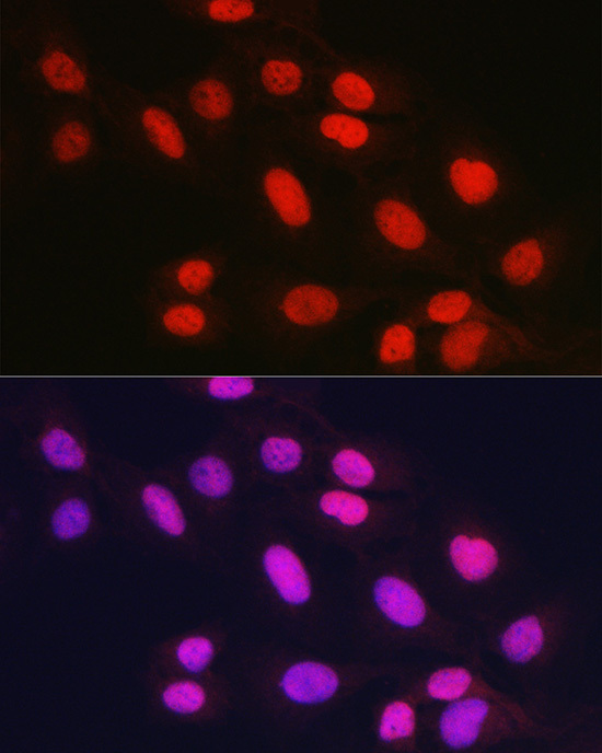
- Immunofluorescence analysis of U2OS cells using Rad21 rabbit polyclonal antibody (STJ11101535) at dilution of 1:150 (40x lens). Blue: DAPI for nuclear staining.
- Sample type
- NA
- Validation comment
- NA
- Primary Ab dilution
- NA
- Other comments
- NA
- Secondary Ab
- NA
- Secondary Ab dilution
- NA
- Protocol
- NA
Supportive validation
- Submitted by
- St John's Laboratory (provider)
- Main image
- Experimental details
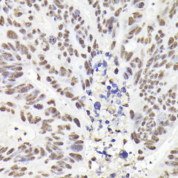
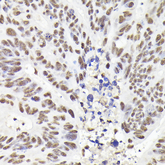
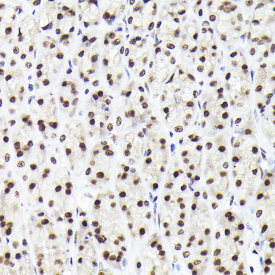
- Immunohistochemistry of paraffin-embedded human colon carcinoma using Rad21 rabbit polyclonal antibody (STJ11101535) at dilution of 1:200 (40x lens). Perform high pressure antigen retrieval with 10 mM citrate buffer pH 6. 0 before commencing with immunohistochemistry staining protocol.
- Sample type
- NA
- Validation comment
- NA
- Primary Ab dilution
- NA
- Other comments
- NA
- Secondary Ab
- NA
- Secondary Ab dilution
- NA
- Protocol
- NA
Supportive validation
- Submitted by
- St John's Laboratory (provider)
- Main image
- Experimental details
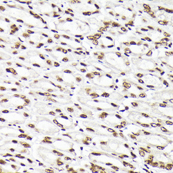
- Immunohistochemistry of paraffin-embedded mouse stomach using Rad21 rabbit polyclonal antibody (STJ11101535) at dilution of 1:200 (40x lens). Perform high pressure antigen retrieval with 10 mM citrate buffer pH 6. 0 before commencing with immunohistochemistry staining protocol.
- Sample type
- NA
- Validation comment
- NA
- Primary Ab dilution
- NA
- Other comments
- NA
- Secondary Ab
- NA
- Secondary Ab dilution
- NA
- Protocol
- NA

Supportive validation
- Submitted by
- St John's Laboratory (provider)
- Main image
- Experimental details
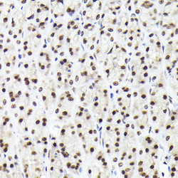
- Immunohistochemistry of paraffin-embedded rat stomach using Rad21 rabbit polyclonal antibody (STJ11101535) at dilution of 1:200 (40x lens). Perform high pressure antigen retrieval with 10 mM citrate buffer pH 6. 0 before commencing with immunohistochemistry staining protocol.
- Sample type
- NA
- Validation comment
- NA
- Primary Ab dilution
- NA
- Other comments
- NA
- Secondary Ab
- NA
- Secondary Ab dilution
- NA
- Protocol
- NA
Supportive validation
Supportive validation
- Submitted by
- St John's Laboratory (provider)
- Main image
- Experimental details
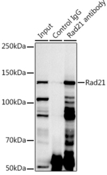
- Immunoprecipitation analysis of 300ug extracts of Jurkat cells using 3ug Rad21 antibody (STJ11101535). Western blot was performed from the immunoprecipitate using Rad21 antibody (STJ11101535) at a dilition of 1:1000.
- Sample type
- NA
- Validation comment
- NA
- Primary Ab dilution
- NA
- Other comments
- NA
- Secondary Ab
- NA
- Secondary Ab dilution
- NA
- Protocol
- NA

Supportive validation
- Submitted by
- St John's Laboratory (provider)
- Main image
- Experimental details
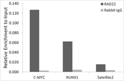
- Chromatin immunoprecipitation analysis of extracts of A-549 cells, using Rad21 antibody (STJ11101535) and rabbit IgG. The amount of immunoprecipitated DNA was checked by quantitative PCR. Histogram was constructed by the ratios of the immunoprecipitated DNA to the input.
- Sample type
- NA
- Validation comment
- NA
- Primary Ab dilution
- NA
- Other comments
- NA
- Secondary Ab
- NA
- Secondary Ab dilution
- NA
- Protocol
- NA
