 Explore
 Validate
 Learn
Learn Western blot
Antibody data
- Antibody Data
- Antigen structure
- References [0]
- Comments [0]
- Validations
- Western blot [7]
- Immunocytochemistry [2]
- Immunohistochemistry [5]
- Chromatin Immunoprecipitation [1]
Submit
Validation data
Reference
Comment
Report error
- Product number
- PA5-28344 - Provider product page

- Provider
- Invitrogen Antibodies
- Product name
- RAD21 Polyclonal Antibody
- Antibody type
- Polyclonal
- Antigen
- Recombinant protein fragment
- Description
- Recommended positive controls: K562, THP-1, HL-60, NIH-3T3, JC, BCL-1.
- Concentration
- 0.44 mg/mL
No comments: Submit comment
Supportive validation
- Submitted by
- Invitrogen Antibodies (provider)
- Main image
- Experimental details
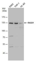
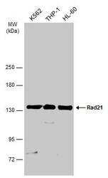
- Western blot analysis of RAD21 using Various whole cell extracts (30 µg). Samples were loaded onto a 7.5% SDS-PAGE gel and probed with a RAD21 polyclonal antibody (Product # PA5-28344) at a dilution of 1:1000.
- Submitted by
- Invitrogen Antibodies (provider)
- Main image
- Experimental details
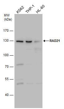
- Western Blot analysis of RAD21 was performed by separating 30 µg of various whole cell extracts by 5% SDS-PAGE. Proteins were transferred to a membrane and probed with a RAD21 Polyclonal Antibody (Product # PA5-28344) at a dilution of 1:1000 and a HRP-conjugated anti-rabbit IgG secondary antibody.
- Submitted by
- Invitrogen Antibodies (provider)
- Main image
- Experimental details
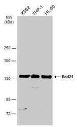
- Western Blot analysis of RAD21 was performed by separating 30 µg of various whole cell extracts by 5% SDS-PAGE. Proteins were transferred to a membrane and probed with a RAD21 Polyclonal Antibody (Product # PA5-28344) at a dilution of 1:1000 and a HRP-conjugated anti-rabbit IgG secondary antibody.

- Submitted by
- Invitrogen Antibodies (provider)
- Main image
- Experimental details
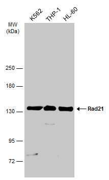
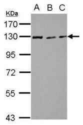
- Western Blot using RAD21 Polyclonal Antibody (Product # PA5-28344). Sample (30 µg of whole cell lysate). Lane A: NIH-3T3. Lane B: JC. Lane C: BCL-1. 7.5% SDS PAGE. RAD21 Polyclonal Antibody (Product # PA5-28344) diluted at 1:3,000. The HRP-conjugated anti-rabbit IgG antibody was used to detect the primary antibody.

- Submitted by
- Invitrogen Antibodies (provider)
- Main image
- Experimental details
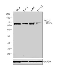
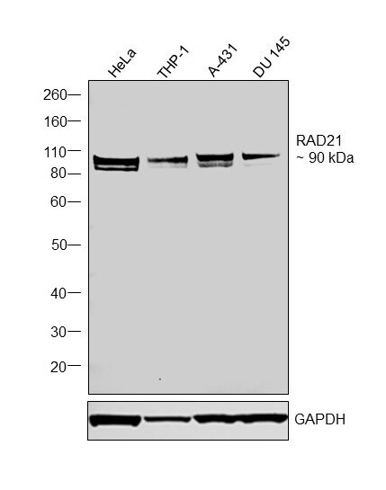
- Western blot was performed using Anti-RAD21 Polyclonal Antibody (Product # PA5-28344) and a 90 kDa band corresponding to RAD21 was observed across tested cell lines. Nuclear enriched extracts (40 µg lysate) of HeLa (Lane 1), THP-1 (Lane 2), A-431 (Lane 3), DU 145 (Lane 4) were electrophoresed using NuPAGE™ 4-12% Bis-Tris Protein Gel (Product # NP0321BOX). Resolved proteins were then transferred onto a nitrocellulose membrane (Product # IB23001) by iBlot® 2 Dry Blotting System (Product # IB21001). The blot was probed with the primary antibody (1:1000 dilution) and detected by chemiluminescence with Goat anti-Rabbit IgG (H+L) Superclonal™ Recombinant Secondary Antibody, HRP (Product # A27036,1:20000 dilution) using the iBright™ FL1500 Imaging System (Product # A44115). Chemiluminescent detection was performed using SuperSignal™ West Pico PLUS Chemiluminescent Substrate (Product # 34580).
- Submitted by
- Invitrogen Antibodies (provider)
- Main image

- Experimental details
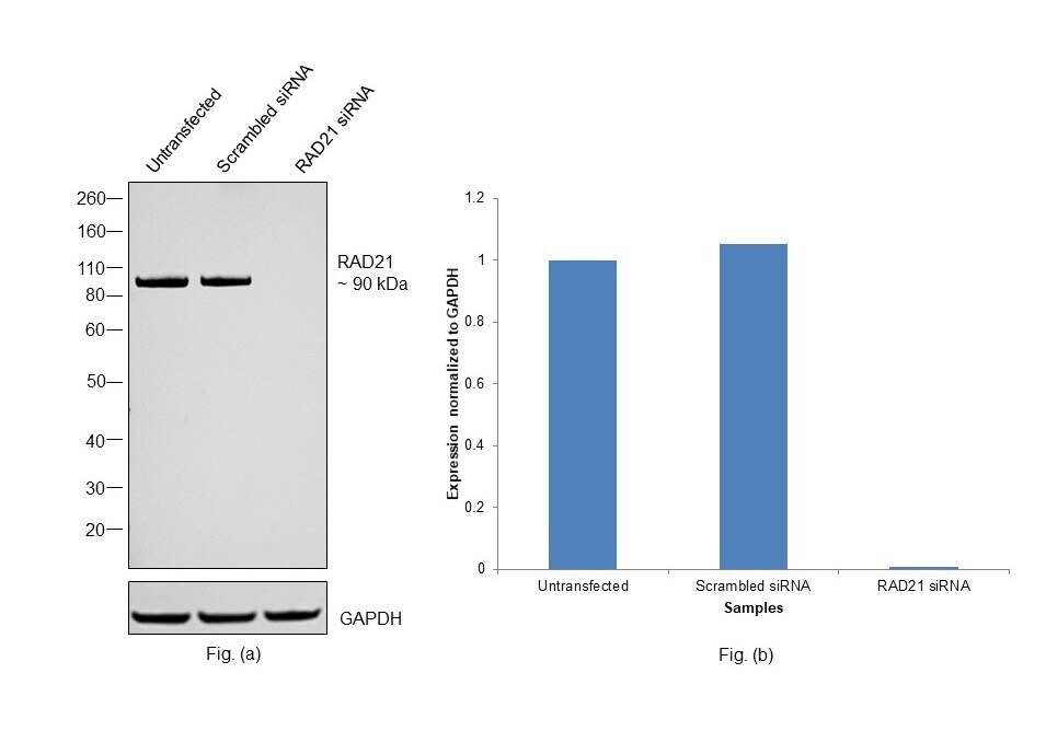
- Knockdown of RAD21 was achieved by transfecting HeLa with RAD21 specific siRNAs (Silencer® select Product # s531211, s531212). Western blot analysis (Fig. a) was performed using Nuclear enriched extracts from the RAD21 knockdown cells (lane 3), non-targeting scrambled siRNA transfected cells (lane 2) and untransfected cells (lane 1). The blot was probed with RAD21 Polyclonal Antibody (Product # PA5-28344, 1:1000 dilution) and Goat anti-Rabbit IgG (H+L) Superclonal™ Recombinant Secondary Antibody, HRP (Product # A27036, 1:20000 dilution). Densitometric analysis of this western blot is shown in histogram (Fig. b). Decrease in signal upon siRNA mediated knock down confirms that antibody is specific to RAD21.
- Submitted by
- Invitrogen Antibodies (provider)
- Main image
- Experimental details
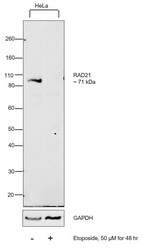
- Western blot using RAD21 Polyclonal Antibody (Product # PA5-28344) and a band at ~90 kDa corresponding to RAD21 was decreased upon etoposide treatment in HeLa. Modified whole cell extracts (1% SDS) (30 µg lysate) of HeLa (Lane 1), HeLa treated with 50µM Etoposide for 48 hrs (Lane 2), were electrophoresed using Novex® NuPAGE® 4-12 % Bis-Tris gel (Product # NP0322BOX). Resolved proteins were then transferred onto a nitrocellulose membrane (Product # IB23001) by iBlot® 2 Dry Blotting System (Product # IB21001). The blot was probed with the primary antibody (1:1000 dilution) and detected by chemiluminescence with Goat anti-Rabbit IgG (H+L) Superclonal™ Recombinant Secondary Antibody, HRP (Product # A27036, 1:4000 dilution) using the iBright FL 1000 (Product # A32752). Chemiluminescent detection was performed using Novex® ECL Chemiluminescent Substrate Reagent Kit (Product # WP20005). Altered expression of proteins upon cell treatment demonstrates antibody specificity.

Supportive validation
- Submitted by
- Invitrogen Antibodies (provider)
- Main image
- Experimental details
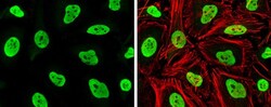
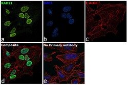
- Immunocytochemistry-Immunofluorescence analysis of RAD21 was performed in HeLa cells fixed in 4% paraformaldehyde at RT for 15 min. Green: RAD21 Polyclonal Antibody (Product # PA5-28344) diluted at 1:1000. Red: phalloidin, a cytoskeleton marker.
- Submitted by
- Invitrogen Antibodies (provider)
- Main image
- Experimental details
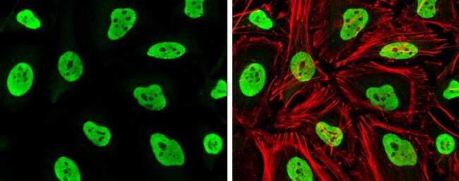
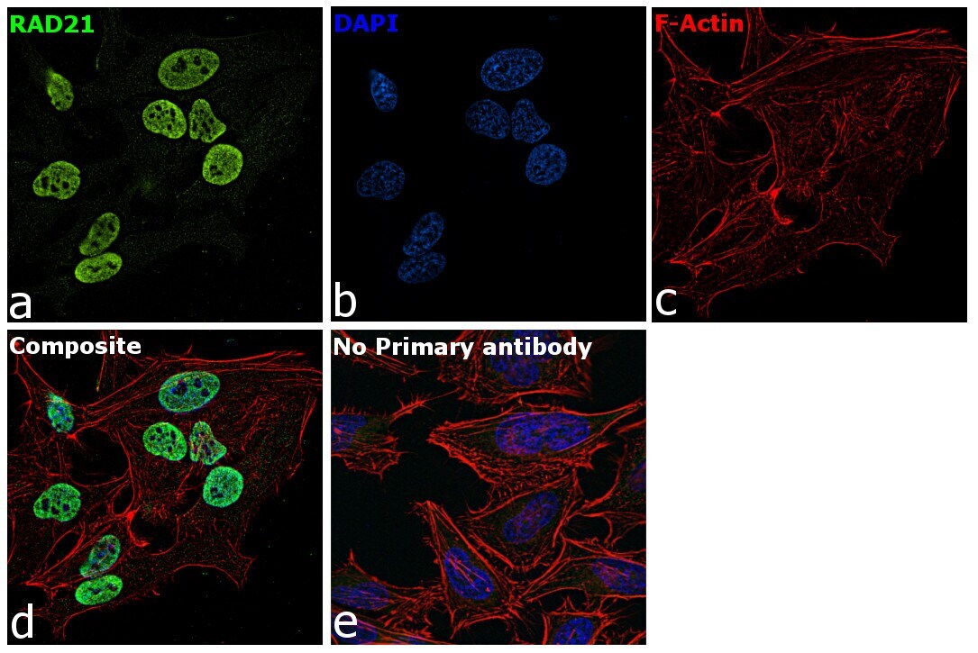
- Immunofluorescence analysis of RAD21 was performed using 70% confluent log phase HeLa cells. The cells were fixed with 4% paraformaldehyde for 10 minutes, permeabilized with 0.1% Triton™ X-100 for 15 minutes, and blocked with 2% BSA for 1 hour at room temperature. The cells were labeled with RAD21 Polyclonal Antibody (Product # PA5-28344) at 5 µg/mL in 0.1% BSA, incubated at 4 degree Celsius overnight and then with Goat anti-Rabbit IgG (H+L) Highly Cross-Adsorbed Secondary Antibody, Alexa Fluor Plus 488 (Product # A32731) at a dilution of 1:2000 for 45 minutes at room temperature (Panel a: green). Nuclei (Panel b: blue) were stained with ProLong™ Diamond Antifade Mountant with DAPI (Product # P36962). F-actin (Panel c: red) was stained with Rhodamine Phalloidin (Product # R415, 1:300). Panel d represents the merged image showing nuclear localization in HeLa cells. Panel e represents control cells with no primary antibody to assess background. The images were captured at 60X magnification.
Supportive validation
- Submitted by
- Invitrogen Antibodies (provider)
- Main image

- Experimental details
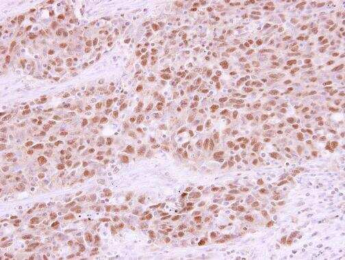
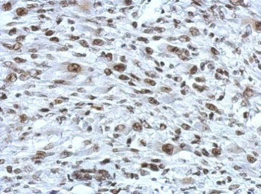
- Immunohistochemistry (Paraffin) analysis of RAD21 was performed in paraffin-embedded human lung papillary adenocarcinoma tissue using RAD21 Polyclonal Antibody (Product # PA5-28344) at a dilution of 1:250.
- Submitted by
- Invitrogen Antibodies (provider)
- Main image
- Experimental details
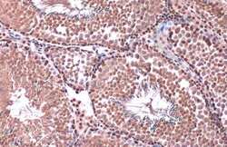
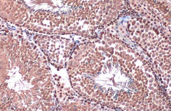
- Immunohistochemistry (Paraffin) analysis of RAD21 was performed in paraffin-embedded mouse testis tissue using RAD21 Polyclonal Antibody (Product # PA5-28344) at a dilution of 1:500. Antigen Retrieval: Citrate buffer, pH 6.0, 15 min.
- Submitted by
- Invitrogen Antibodies (provider)
- Main image
- Experimental details
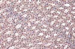
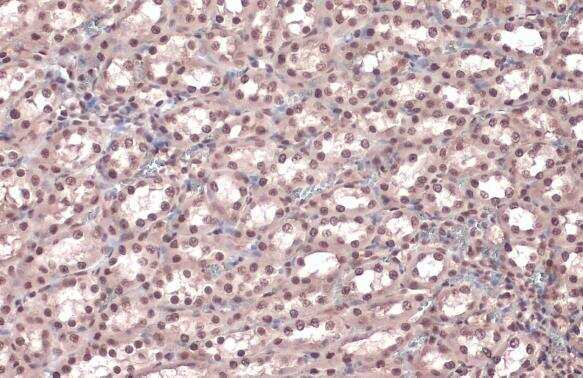
- Immunohistochemistry (Paraffin) analysis of RAD21 was performed in paraffin-embedded rat kidney tissue using RAD21 Polyclonal Antibody (Product # PA5-28344) at a dilution of 1:500. Antigen Retrieval: Citrate buffer, pH 6.0, 15 min.
- Submitted by
- Invitrogen Antibodies (provider)
- Main image
- Experimental details
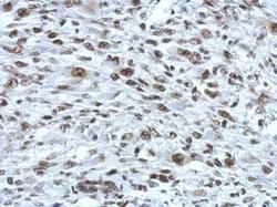
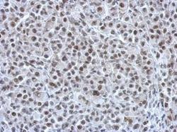
- Immunohistochemical analysis of paraffin-embedded C2C12 xenograft, using RAD21 (Product # PA5-28344) antibody at 1:750 dilution. Antigen Retrieval: EDTA based buffer, pH 8.0, 15 min.
- Submitted by
- Invitrogen Antibodies (provider)
- Main image
- Experimental details
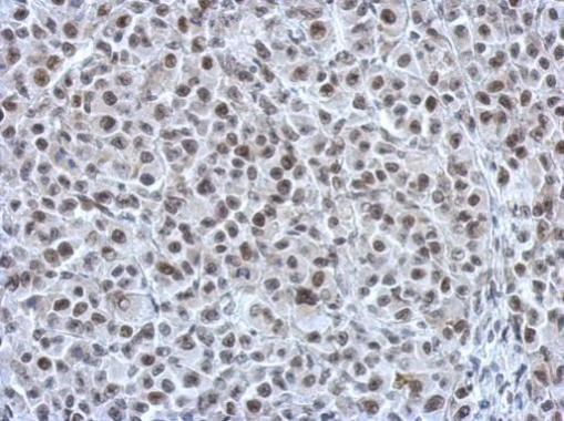
- Immunohistochemical analysis of paraffin-embedded HBL435 xenograft, using RAD21 (Product # PA5-28344) antibody at 1:750 dilution. Antigen Retrieval: EDTA based buffer, pH 8.0, 15 min.
Supportive validation
- Submitted by
- Invitrogen Antibodies (provider)
- Main image
- Experimental details
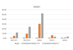
- Chromatin Immunoprecipitation (ChIP) assay of endogenous RAD21 protein using RAD21 Antibody: ChIP was performed using RAD21 Polyclonal Antibody (Product # PA5-28344, 5 µg) on sheared chromatin from MCF7 and MCF7 cells treated with Estradiol (100 nM, 45 minutes) using the MAGnify ChIP System kit (Product # 49-2024). Normal Rabbit IgG was used as a negative IP control. The purified DNA was analyzed by qPCR using primers binding to MYC promotor regions (P1 and P2), ERE2 (active) and MYC1 (-394kb of myc TSS), SAT alpha (Inactive). Data is presented as fold enrichment of the antibody signal versus the negative control IgG using the comparative CT method.
