Explore
 Validate
 Learn
Learn Western blot
 Immunocytochemistry
Antibody data
- Antibody Data
- Antigen structure
- References [8]
- Comments [0]
- Validations
- Immunocytochemistry [4]
- Other assay [2]
Submit
Validation data
Reference
Comment
Report error
- Product number
- PA1-330 - Provider product page

- Provider
- Invitrogen Antibodies
- Product name
- LXR alpha Polyclonal Antibody
- Antibody type
- Polyclonal
- Antigen
- Synthetic peptide
- Description
- PA1-330 detects recombinant human liver X receptor (LXR) alpha. This antibody does not detect human recombinant LXR beta protein. PA1-330 has been successfully used in ICC/IF and Western blot procedures. By Western blot, this antibody detects an ~64 kDa protein representing recombinant human LXR alpha. The PA1-330 immunogen is a synthetic peptide corresponding to residues C D(318) F S Y N R E D F A K A(329) of human LXR alpha. The PA1-330 immunizing peptide (Cat. # PEP-114) is available for use in neutralization and control experiments.
- Reactivity
- Human
- Host
- Rabbit
- Isotype
- IgG
- Vial size
- 200 μg
- Concentration
- 1 mg/mL
- Storage
- -20°C, Avoid Freeze/Thaw Cycles
Submitted references Administration of GDF3 Into Septic Mice Improves Survival via Enhancing LXRα-Mediated Macrophage Phagocytosis.
Sectm1a deficiency aggravates inflammation-triggered cardiac dysfunction through disruption of LXRα signalling in macrophages.
Macrophage miR-34a Is a Key Regulator of Cholesterol Efflux and Atherosclerosis.
27-hydroxycholesterol decreases cell proliferation in colon cancer cell lines.
Liver X receptor is a therapeutic target in collagen-induced arthritis.
Endotoxin down-regulates ABCG5 and ABCG8 in mouse liver and ABCA1 and ABCG1 in J774 murine macrophages: differential role of LXR.
Oxysterol-activated LXRalpha/RXR induces hSR-BI-promoter activity in hepatoma cells and preadipocytes.
Oxysterol-activated LXRalpha/RXR induces hSR-BI-promoter activity in hepatoma cells and preadipocytes.
Wang P, Mu X, Zhao H, Li Y, Wang L, Wolfe V, Cui SN, Wang X, Peng T, Zingarelli B, Wang C, Fan GC
Frontiers in immunology 2021;12:647070
Frontiers in immunology 2021;12:647070
Sectm1a deficiency aggravates inflammation-triggered cardiac dysfunction through disruption of LXRα signalling in macrophages.
Li Y, Deng S, Wang X, Huang W, Chen J, Robbins N, Mu X, Essandoh K, Peng T, Jegga AG, Rubinstein J, Adams DE, Wang Y, Peng J, Fan GC
Cardiovascular research 2021 Feb 22;117(3):890-902
Cardiovascular research 2021 Feb 22;117(3):890-902
Macrophage miR-34a Is a Key Regulator of Cholesterol Efflux and Atherosclerosis.
Xu Y, Xu Y, Zhu Y, Sun H, Juguilon C, Li F, Fan D, Yin L, Zhang Y
Molecular therapy : the journal of the American Society of Gene Therapy 2020 Jan 8;28(1):202-216
Molecular therapy : the journal of the American Society of Gene Therapy 2020 Jan 8;28(1):202-216
27-hydroxycholesterol decreases cell proliferation in colon cancer cell lines.
Warns J, Marwarha G, Freking N, Ghribi O
Biochimie 2018 Oct;153:171-180
Biochimie 2018 Oct;153:171-180
Liver X receptor is a therapeutic target in collagen-induced arthritis.
Chintalacharuvu SR, Sandusky GE, Burris TP, Burmer GC, Nagpal S
Arthritis and rheumatism 2007 Apr;56(4):1365-7
Arthritis and rheumatism 2007 Apr;56(4):1365-7
Endotoxin down-regulates ABCG5 and ABCG8 in mouse liver and ABCA1 and ABCG1 in J774 murine macrophages: differential role of LXR.
Khovidhunkit W, Moser AH, Shigenaga JK, Grunfeld C, Feingold KR
Journal of lipid research 2003 Sep;44(9):1728-36
Journal of lipid research 2003 Sep;44(9):1728-36
Oxysterol-activated LXRalpha/RXR induces hSR-BI-promoter activity in hepatoma cells and preadipocytes.
Malerød L, Juvet LK, Hanssen-Bauer A, Eskild W, Berg T
Biochemical and biophysical research communications 2002 Dec 20;299(5):916-23
Biochemical and biophysical research communications 2002 Dec 20;299(5):916-23
No comments: Submit comment
Supportive validation
- Submitted by
- Invitrogen Antibodies (provider)
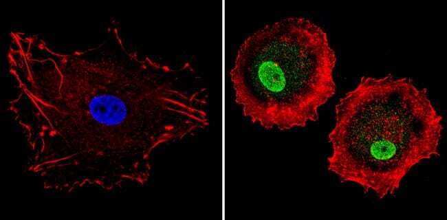
- Main image
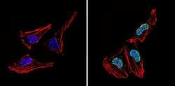
- Experimental details
- Immunofluorescent analysis of LXR alpha (green) showing staining in the nucleus of Hela cells (right) compared to a negative control without primary antibody (left). Formalin-fixed cells were permeabilized with 0.1% Triton X-100 in TBS for 5-10 minutes and blocked with 3% BSA-PBS for 30 minutes at room temperature. Cells were probed with a LXR alpha polyclonal antibody (Product # PA1-330) in 3% BSA-PBS at a dilution of 1:300 and incubated overnight at 4ºC in a humidified chamber. Cells were washed with PBST and incubated with a DyLight-conjugated secondary antibody in PBS at room temperature in the dark. F-actin (red) was stained with a fluorescent red phalloidin and nuclei (blue) were stained with Hoechst or DAPI. Images were taken at a magnification of 60x.
- Submitted by
- Invitrogen Antibodies (provider)
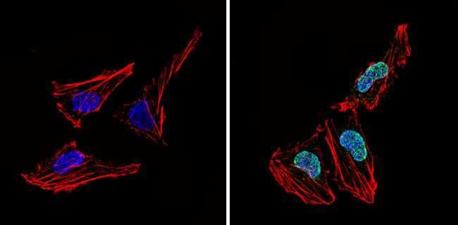
- Main image
- Experimental details
- Immunofluorescent analysis of LXR alpha (green) showing staining in the nucleus of MCF-7 cells (right) compared to a negative control without primary antibody (left). Formalin-fixed cells were permeabilized with 0.1% Triton X-100 in TBS for 5-10 minutes and blocked with 3% BSA-PBS for 30 minutes at room temperature. Cells were probed with a LXR alpha polyclonal antibody (Product # PA1-330) in 3% BSA-PBS at a dilution of 1:300 and incubated overnight at 4ºC in a humidified chamber. Cells were washed with PBST and incubated with a DyLight-conjugated secondary antibody in PBS at room temperature in the dark. F-actin (red) was stained with a fluorescent red phalloidin and nuclei (blue) were stained with Hoechst or DAPI. Images were taken at a magnification of 60x.
- Submitted by
- Invitrogen Antibodies (provider)
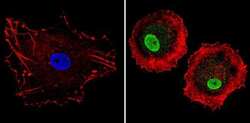
- Main image
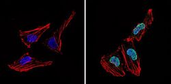
- Experimental details
- Immunofluorescent analysis of LXR alpha (green) showing staining in the nucleus of Hela cells (right) compared to a negative control without primary antibody (left). Formalin-fixed cells were permeabilized with 0.1% Triton X-100 in TBS for 5-10 minutes and blocked with 3% BSA-PBS for 30 minutes at room temperature. Cells were probed with a LXR alpha polyclonal antibody (Product # PA1-330) in 3% BSA-PBS at a dilution of 1:300 and incubated overnight at 4ºC in a humidified chamber. Cells were washed with PBST and incubated with a DyLight-conjugated secondary antibody in PBS at room temperature in the dark. F-actin (red) was stained with a fluorescent red phalloidin and nuclei (blue) were stained with Hoechst or DAPI. Images were taken at a magnification of 60x.
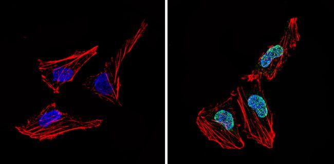
- Submitted by
- Invitrogen Antibodies (provider)
- Main image

- Experimental details
- Immunofluorescent analysis of LXR alpha (green) showing staining in the nucleus of MCF-7 cells (right) compared to a negative control without primary antibody (left). Formalin-fixed cells were permeabilized with 0.1% Triton X-100 in TBS for 5-10 minutes and blocked with 3% BSA-PBS for 30 minutes at room temperature. Cells were probed with a LXR alpha polyclonal antibody (Product # PA1-330) in 3% BSA-PBS at a dilution of 1:300 and incubated overnight at 4ºC in a humidified chamber. Cells were washed with PBST and incubated with a DyLight-conjugated secondary antibody in PBS at room temperature in the dark. F-actin (red) was stained with a fluorescent red phalloidin and nuclei (blue) were stained with Hoechst or DAPI. Images were taken at a magnification of 60x.

Supportive validation
- Submitted by
- Invitrogen Antibodies (provider)
- Main image
- Experimental details
- NULL
- Submitted by
- Invitrogen Antibodies (provider)
- Main image
- Experimental details
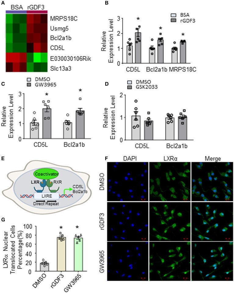
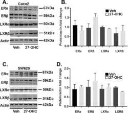
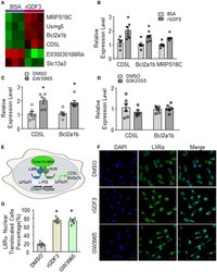
- Figure 5 Gene expression profile in BMDMs treated with rGDF3 by high-throughput RNA sequencing. (A) Heatmap of the differentially expressed genes from BMDMs treated with BSA or rGDF3 (20 ng/mL) ( n = 3). (B) The altered expression of CD5L, Bcl2a1b, and MRPS18C gene in rGDF3-treated BMDMs was validated by qRT-PCR ( n = 6). (C) Expression of CD5L and Bcl2a1b in GW3965 (1 mumol/L)-treated BMDMs was determined by qRT-PCR ( n = 6) at 18 h post-treatment. (D) Expression of CD5L and Bcl2a1b in BMDMs were measured at 18 h post-GSK2033 (2 mumol/L) treatment ( n = 6). (E) Graphic scheme of the LXRalpha-CD5L signal cascade in macrophages. Representative images (F) and quantification (G) of immunofluorescence staining for LXRalpha (green) in BMDMs at 18 h after rGDF3 (20 ng/mL) or GW3965 (1 mumol/L) treatment (Scale bar, 10 mum; n = 5). Data are representative of two (B-D,F,G) independent experiments. All results are shown as mean +- SEM and analyzed by Student's t -test (* p < 0.05).