Explore
 Validate
 Learn
Learn Western blot
 Immunocytochemistry
Antibody data
- Antibody Data
- Antigen structure
- References [5]
- Comments [0]
- Validations
- Western blot [2]
- Flow cytometry [1]
Submit
Validation data
Reference
Comment
Report error
- Product number
- NB300-612 - Provider product page

- Provider
- Novus Biologicals
- Proper citation
- Novus Cat#NB300-612, RRID:AB_11046430
- Product name
- Rabbit Polyclonal LXR alpha/NR1H3 Antibody
- Antibody type
- Polyclonal
- Description
- Immunogen affinity purified. Detects recombinant human LXR alpha. This does not detect recombinant human LXR beta.
- Reactivity
- Human, Mouse
- Host
- Rabbit
- Isotype
- IgG
- Vial size
- 200 ug
- Concentration
- 1 mg/ml
- Storage
- Store at -20C. Avoid freeze-thaw cycles.
Submitted references PNPLA3 I148M Variant Impairs Liver X Receptor Signaling and Cholesterol Homeostasis in Human Hepatic Stellate Cells.
Activation of a prometastatic gene expression program in hypoxic neuroblastoma cells.
Suppression of 2,3-oxidosqualene cyclase by high fat diet contributes to liver X receptor-alpha-mediated improvement of hepatic lipid profile.
Discovery and implementation of transcriptional biomarkers of synthetic LXR agonists in peripheral blood cells.
NO-1886 upregulates ATP binding cassette transporter A1 and inhibits diet-induced atherosclerosis in Chinese Bama minipigs.
Bruschi FV, Claudel T, Tardelli M, Starlinger P, Marra F, Trauner M
Hepatology communications 2019 Sep;3(9):1191-1204
Hepatology communications 2019 Sep;3(9):1191-1204
Activation of a prometastatic gene expression program in hypoxic neuroblastoma cells.
Poomthavorn P, Wong SH, Higgins S, Werther GA, Russo VC
Endocrine-related cancer 2009 Sep;16(3):991-1004
Endocrine-related cancer 2009 Sep;16(3):991-1004
Suppression of 2,3-oxidosqualene cyclase by high fat diet contributes to liver X receptor-alpha-mediated improvement of hepatic lipid profile.
Dang H, Liu Y, Pang W, Li C, Wang N, Shyy JY, Zhu Y
The Journal of biological chemistry 2009 Mar 6;284(10):6218-26
The Journal of biological chemistry 2009 Mar 6;284(10):6218-26
Discovery and implementation of transcriptional biomarkers of synthetic LXR agonists in peripheral blood cells.
DiBlasio-Smith EA, Arai M, Quinet EM, Evans MJ, Kornaga T, Basso MD, Chen L, Feingold I, Halpern AR, Liu QY, Nambi P, Savio D, Wang S, Mounts WM, Isler JA, Slager AM, Burczynski ME, Dorner AJ, LaVallie ER
Journal of translational medicine 2008 Oct 16;6:59
Journal of translational medicine 2008 Oct 16;6:59
NO-1886 upregulates ATP binding cassette transporter A1 and inhibits diet-induced atherosclerosis in Chinese Bama minipigs.
Zhang C, Yin W, Liao D, Huang L, Tang C, Tsutsumi K, Wang Z, Liu Y, Li Q, Hou H, Cai M, Xiao J
Journal of lipid research 2006 Sep;47(9):2055-63
Journal of lipid research 2006 Sep;47(9):2055-63
No comments: Submit comment
Supportive validation
- Submitted by
- Novus Biologicals (provider)
- Main image
- Experimental details
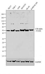
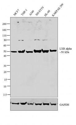
- Western Blot: LXR alpha/NR1H3 Antibody [NB300-612] - analysis was performed on nuclear enriched extracts (30 ug lysate) of MCF7 (Lane 1), THP-1 (Lane 2), A549 (Lane 3), SH-SY5Y (Lane 4), HL-60 (Lane 5) and KARPAS 299 (Lane 6). The blot was probed with Rabbit Anti-LXR alpha Polyclonal Antibody (2ug/ml) and detected by chemiluminescence using Goat anti-Rabbit IgG (H+L) Secondary Antibody, HRP conjugate (0.25 ug/ml, 1:4000 dilution). A 50 kDa band corresponding to LXR alpha was observed across the cell lines tested. Known quantity of protein samples was electrophoresed using 4-12 % Bis-Tris gel, Electrophoresis System and Sharp Pre-Stained Protein Standard. Resolved proteins were then transferred onto a nitrocellulose membrane with Blot 2 Dry Blotting System. The membrane was probed with the relevant primary and secondary Antibody following blocking with 5 % skimmed milk. Chemiluminescent detection was performed using ECL Chemiluminescent Substrate Reagent Kit.
- Submitted by
- Novus Biologicals (provider)
- Main image
- Experimental details
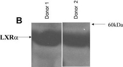
- Western Blot: LXR alpha/NR1H3 Antibody [NB300-612] - LXRs are expressed in peripheral blood cells. LXR alpha/NR1H3 protein levels in protein extracts from PBMCs from these same donors were detected by Western blotting using rabbit anti-human LXR alpha/NR1H3 polyclonal antisera. Image collected and cropped by CiteAb from the following publication (http://translational-medicine.biomedcentral.com/articles/10.1186/1479-5876-6-59), licensed under a CC-BY licence.

Supportive validation
- Submitted by
- Novus Biologicals (provider)
- Main image
- Experimental details
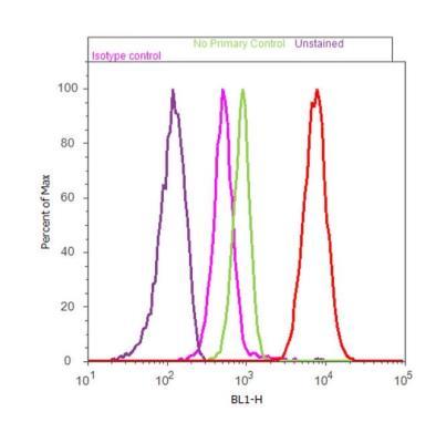
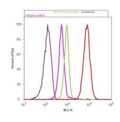
- Flow Cytometry: LXR alpha/NR1H3 Antibody [NB300-612] - analysis of LXR alpha was done on HeLa. Cells were fixed with 70% ethanol for 10 minutes, permeabilized with 0.25% Triton X-100 for 20 minutes, and blocked with 2.5% BSA for 30 minutes at room temperature. Cells were labeled with LXR alpha Rabbit Polyclonal Antibody (PA1330, red histogram) or with rabbit isotype control (pink histogram) at 3-5 ug/million cells in 2.5% BSA. After incubation at room temperature for 2 hours, the cells were labeled with Alexa Fluor 488 Goat Anti-Rabbit Secondary Antibody (A11008) at a dilution of 1:400 for 30 minutes at room temperature. The representative 10,000 cells were acquired and analyzed for each sample using an Acoustic Focusing Cytometer. The purple histogram represents unstained control cells and the green histogram represents no-primary-antibody control.