Explore
 Validate
 Learn
Learn Western blot
 Immunoprecipitation
 Other assay
Antibody data
- Antibody Data
- Antigen structure
- References [19]
- Comments [0]
- Validations
- Other assay [8]
Submit
Validation data
Reference
Comment
Report error
- Product number
- 34-6200 - Provider product page

- Provider
- Invitrogen Antibodies
- Product name
- MAG Polyclonal Antibody
- Antibody type
- Polyclonal
- Antigen
- Other
- Reactivity
- Human, Mouse, Rat
- Host
- Rabbit
- Isotype
- IgG
- Vial size
- 100 μg
- Concentration
- 0.25 mg/mL
- Storage
- -20°C
Submitted references Therapeutic advantages of combined gene/cell therapy strategies in a murine model of GM2 gangliosidosis.
The Etv1/Er81 transcription factor coordinates myelination-related genes to regulate Schwann cell differentiation and myelination.
Clemastine improves electrophysiologic and histomorphometric changes through promoting myelin repair in a murine model of compression neuropathy.
Non-canonical Targets of HIF1a Impair Oligodendrocyte Progenitor Cell Function.
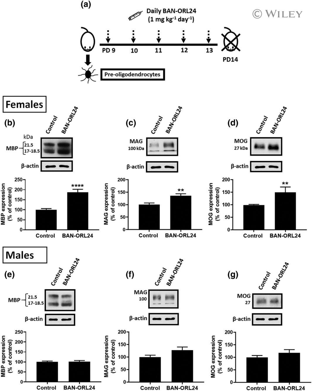
Endogenous opioid peptides and brain development: Endomorphin-1 and Nociceptin play a sex-specific role in the control of oligodendrocyte maturation and brain myelination.
Deletion of Calcineurin in Schwann Cells Does Not Affect Developmental Myelination, But Reduces Autophagy and Delays Myelin Clearance after Peripheral Nerve Injury.
Schwann cells, but not Oligodendrocytes, Depend Strictly on Dynamin 2 Function.
Neuregulin 1 type III improves peripheral nerve myelination in a mouse model of congenital hypomyelinating neuropathy.
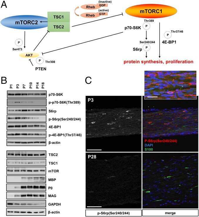
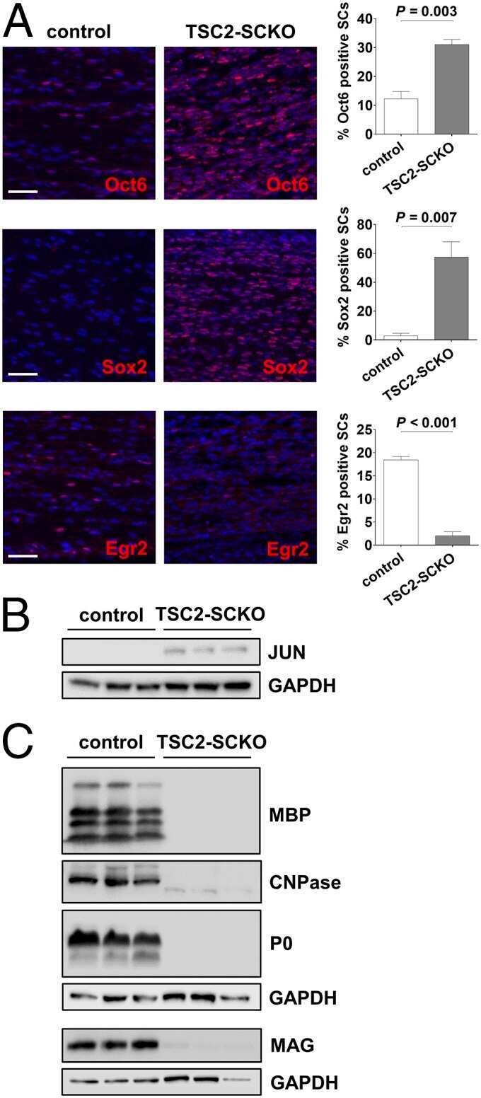
mTORC1 promotes proliferation of immature Schwann cells and myelin growth of differentiated Schwann cells.
LINGO-1 Regulates Oligodendrocyte Differentiation through the Cytoplasmic Gelsolin Signaling Pathway.
The integrated stress response in hypoxia-induced diffuse white matter injury.
Defects of Lipid Synthesis Are Linked to the Age-Dependent Demyelination Caused by Lamin B1 Overexpression.
Very large G protein-coupled receptor 1 regulates myelin-associated glycoprotein via Gαs/Gαq-mediated protein kinases A/C.
Upregulation of axon guidance molecules in the adult central nervous system of Nogo-A knockout mice restricts neuronal growth and regeneration.
Characterizing Mystery Cell Lines: Student-driven Research Projects in an Undergraduate Neuroscience Laboratory Course.
Conduction block in PMP22 deficiency.
Mutation of FIG4 causes a rapidly progressive, asymmetric neuronal degeneration.
Growth-associated gene and protein expression in the region of axonal sprouting in the aged brain after stroke.
Histone modifications affect timing of oligodendrocyte progenitor differentiation in the developing rat brain.
Sala D, Ornaghi F, Morena F, Argentati C, Valsecchi M, Alberizzi V, Di Guardo R, Bolino A, Aureli M, Martino S, Gritti A
Molecular therapy. Methods & clinical development 2022 Jun 9;25:170-189
Molecular therapy. Methods & clinical development 2022 Jun 9;25:170-189
The Etv1/Er81 transcription factor coordinates myelination-related genes to regulate Schwann cell differentiation and myelination.
Askar P, Xu J, Hu J, Shangguan J, Sun H, Zhou S, Yang X, Chen G, Su W, Gu Y
Annals of translational medicine 2022 Aug;10(16):875
Annals of translational medicine 2022 Aug;10(16):875
Clemastine improves electrophysiologic and histomorphometric changes through promoting myelin repair in a murine model of compression neuropathy.
Lee JI, Park JW, Lee KJ, Lee DH
Scientific reports 2021 Oct 22;11(1):20886
Scientific reports 2021 Oct 22;11(1):20886
Non-canonical Targets of HIF1a Impair Oligodendrocyte Progenitor Cell Function.
Allan KC, Hu LR, Scavuzzo MA, Morton AR, Gevorgyan AS, Cohn EF, Clayton BLL, Bederman IR, Hung S, Bartels CF, Madhavan M, Tesar PJ
Cell stem cell 2021 Feb 4;28(2):257-272.e11
Cell stem cell 2021 Feb 4;28(2):257-272.e11
Endogenous opioid peptides and brain development: Endomorphin-1 and Nociceptin play a sex-specific role in the control of oligodendrocyte maturation and brain myelination.
Mohamed E, Paisley CE, Meyer LC, Bigbee JW, Sato-Bigbee C
Glia 2020 Jul;68(7):1513-1530
Glia 2020 Jul;68(7):1513-1530
Deletion of Calcineurin in Schwann Cells Does Not Affect Developmental Myelination, But Reduces Autophagy and Delays Myelin Clearance after Peripheral Nerve Injury.
Reed CB, Frick LR, Weaver A, Sidoli M, Schlant E, Feltri ML, Wrabetz L
The Journal of neuroscience : the official journal of the Society for Neuroscience 2020 Aug 5;40(32):6165-6176
The Journal of neuroscience : the official journal of the Society for Neuroscience 2020 Aug 5;40(32):6165-6176
Schwann cells, but not Oligodendrocytes, Depend Strictly on Dynamin 2 Function.
Gerber D, Ghidinelli M, Tinelli E, Somandin C, Gerber J, Pereira JA, Ommer A, Figlia G, Miehe M, Nägeli LG, Suter V, Tadini V, Sidiropoulos PN, Wessig C, Toyka KV, Suter U
eLife 2019 Jan 16;8
eLife 2019 Jan 16;8
Neuregulin 1 type III improves peripheral nerve myelination in a mouse model of congenital hypomyelinating neuropathy.
Belin S, Ornaghi F, Shackleford G, Wang J, Scapin C, Lopez-Anido C, Silvestri N, Robertson N, Williamson C, Ishii A, Taveggia C, Svaren J, Bansal R, Schwab MH, Nave K, Fratta P, D'Antonio M, Poitelon Y, Feltri ML, Wrabetz L
Human molecular genetics 2019 Apr 15;28(8):1260-1273
Human molecular genetics 2019 Apr 15;28(8):1260-1273
mTORC1 promotes proliferation of immature Schwann cells and myelin growth of differentiated Schwann cells.
Beirowski B, Wong KM, Babetto E, Milbrandt J
Proceedings of the National Academy of Sciences of the United States of America 2017 May 23;114(21):E4261-E4270
Proceedings of the National Academy of Sciences of the United States of America 2017 May 23;114(21):E4261-E4270
LINGO-1 Regulates Oligodendrocyte Differentiation through the Cytoplasmic Gelsolin Signaling Pathway.
Shao Z, Lee X, Huang G, Sheng G, Henderson CE, Louvard D, Sohn J, Pepinsky B, Mi S
The Journal of neuroscience : the official journal of the Society for Neuroscience 2017 Mar 22;37(12):3127-3137
The Journal of neuroscience : the official journal of the Society for Neuroscience 2017 Mar 22;37(12):3127-3137
The integrated stress response in hypoxia-induced diffuse white matter injury.
Clayton BL, Huang A, Kunjamma RB, Solanki A, Popko B
The Journal of neuroscience : the official journal of the Society for Neuroscience 2017 Jul 18;37(31):7465-80
The Journal of neuroscience : the official journal of the Society for Neuroscience 2017 Jul 18;37(31):7465-80
Defects of Lipid Synthesis Are Linked to the Age-Dependent Demyelination Caused by Lamin B1 Overexpression.
Rolyan H, Tyurina YY, Hernandez M, Amoscato AA, Sparvero LJ, Nmezi BC, Lu Y, Estécio MR, Lin K, Chen J, He RR, Gong P, Rigatti LH, Dupree J, Bayır H, Kagan VE, Casaccia P, Padiath QS
The Journal of neuroscience : the official journal of the Society for Neuroscience 2015 Aug 26;35(34):12002-17
The Journal of neuroscience : the official journal of the Society for Neuroscience 2015 Aug 26;35(34):12002-17
Very large G protein-coupled receptor 1 regulates myelin-associated glycoprotein via Gαs/Gαq-mediated protein kinases A/C.
Shin D, Lin ST, Fu YH, Ptácek LJ
Proceedings of the National Academy of Sciences of the United States of America 2013 Nov 19;110(47):19101-6
Proceedings of the National Academy of Sciences of the United States of America 2013 Nov 19;110(47):19101-6
Upregulation of axon guidance molecules in the adult central nervous system of Nogo-A knockout mice restricts neuronal growth and regeneration.
Kempf A, Montani L, Petrinovic MM, Schroeter A, Weinmann O, Patrignani A, Schwab ME
The European journal of neuroscience 2013 Dec;38(11):3567-79
The European journal of neuroscience 2013 Dec;38(11):3567-79
Characterizing Mystery Cell Lines: Student-driven Research Projects in an Undergraduate Neuroscience Laboratory Course.
Lemons ML
Journal of undergraduate neuroscience education : JUNE : a publication of FUN, Faculty for Undergraduate Neuroscience 2012 Spring;10(2):A96-A104
Journal of undergraduate neuroscience education : JUNE : a publication of FUN, Faculty for Undergraduate Neuroscience 2012 Spring;10(2):A96-A104
Conduction block in PMP22 deficiency.
Bai Y, Zhang X, Katona I, Saporta MA, Shy ME, O'Malley HA, Isom LL, Suter U, Li J
The Journal of neuroscience : the official journal of the Society for Neuroscience 2010 Jan 13;30(2):600-8
The Journal of neuroscience : the official journal of the Society for Neuroscience 2010 Jan 13;30(2):600-8
Mutation of FIG4 causes a rapidly progressive, asymmetric neuronal degeneration.
Zhang X, Chow CY, Sahenk Z, Shy ME, Meisler MH, Li J
Brain : a journal of neurology 2008 Aug;131(Pt 8):1990-2001
Brain : a journal of neurology 2008 Aug;131(Pt 8):1990-2001
Growth-associated gene and protein expression in the region of axonal sprouting in the aged brain after stroke.
Li S, Carmichael ST
Neurobiology of disease 2006 Aug;23(2):362-73
Neurobiology of disease 2006 Aug;23(2):362-73
Histone modifications affect timing of oligodendrocyte progenitor differentiation in the developing rat brain.
Shen S, Li J, Casaccia-Bonnefil P
The Journal of cell biology 2005 May 23;169(4):577-89
The Journal of cell biology 2005 May 23;169(4):577-89
No comments: Submit comment
Supportive validation
- Submitted by
- Invitrogen Antibodies (provider)
- Main image
- Experimental details
- NULL
- Submitted by
- Invitrogen Antibodies (provider)
- Main image
- Experimental details
- NULL
- Submitted by
- Invitrogen Antibodies (provider)
- Main image
- Experimental details
- NULL
- Submitted by
- Invitrogen Antibodies (provider)
- Main image

- Experimental details
- Administration of an NOR antagonist results in sex-specific acceleration of rat brain myelination. (a) Experimental schedule of BAN-ORL 24 administration: 9-day-old female and male pups were administered the blood brain barrier permeable NOR antagonist BAN-ORL24 (1 mg/kg/day, IP) or vehicle (controls). Animals were sacrificed at postnatal day 14, and the cerebral hemispheres analyzed for the expression of MBP, MAG and MOG, using beta-Actin levels as loading controls. The figure shows representative western blots for the female (b-d) and male (e-g) pups. Bar graphs are the mean +- SEM from a total of at least 9 pups/group/sex from three different litters. **** p < .001, ** p < .006
- Submitted by
- Invitrogen Antibodies (provider)
- Main image
- Experimental details
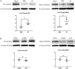
- Figure 4 Representative Western immunoblotting for P0 and MAG protein in compression phase ( a ) and decompression phase ( b ) (P0, myelin protein 0; MAG, myelin associated glycoprotein; Sham, nerves of sham-operated mice; PBS, nerves of untreated mice during compression phase; CF, nerves of mice treated with clemastine during compression phase; d-PBS, nerves of untreated mice after surgical decompression; d-CF, nerves of mice treated with clemastine after surgical decompression). Full-length Western blots are presented in Supplementary Figs. 1 and 2 . Band intensity quantification of P0 and MAG of CF group as fold change of PBS group. Band intensity quantification of P0 and MAG of d-CF group as fold change of d-PBS group. ss-actin was used to normalize the intensity of the bands. Error bars represent standard error of mean. Mann-Whitney U test was conducted (*p < 0.05, and n = 3 or 4 animals per group).

- Submitted by
- Invitrogen Antibodies (provider)
- Main image
- Experimental details
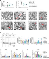
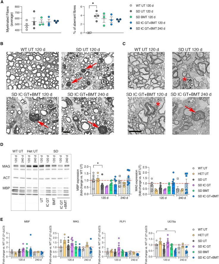
- Analyses of the myelin compartment in treated SD mice (A) Quantification of myelinated fibers and percentage of demyelinated/degenerated (aberrant) fibers among total fibers in SC sections from treated mice (IC GT, BMT, and IC GT + BMT) at 120 days and 240 days and age-matched UT controls (WT and SD). Data are expressed as mean +- SEM; n = 3-4 mice/group; nonparametric one-way ANOVA followed by Dunn's multiple comparisons test, *p < 0.05. (B) Semithin section analysis of lumbar SC of IC GT + BMT-treated SD mice at 120 days and 240 days and UT controls (WT and SD). Red arrows indicate aberrant myelin. Scale bar, 10 mum. (C) Electron microscopy images of optic nerves in treated SD and UT controls at 120 days and 240 days. Red arrows indicate aberrant myelin, and asterisks mark macrophages. Scale bar, 2 mum. (D) Representative western blot analyses and relative quantification showing MBP and MAG protein expression in whole-brain lysate (TEL and CB) of treated mice (IC GT, BMT, and IC GT + BMT) at 120 days and 240 days and age-matched UT controls (WT, Het, and SD). Data are expressed as fold change with respect to the WT (set as 1) after normalization to beta-actin expression. Data represent the mean +- SEM; n = 3-10 mice/group. One-way ANOVA followed by Kruskal-Wallis multiple comparisons test, *p < 0.05. (E) Expression of myelin-related genes ( Mbp , Mag , Plp1 , and Ugt8a ) in whole-brain lysate (TEL and CB) of treated mice (IC GT, BMT, and IC GT + BMT) at 120 days and 240 day
- Submitted by
- Invitrogen Antibodies (provider)
- Main image

- Experimental details
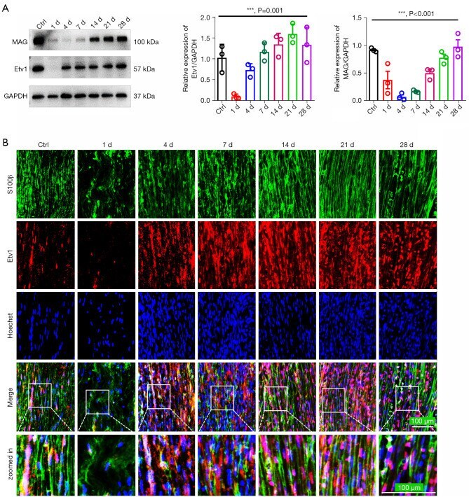
- Etv1 expression and cellular localization in the crushed mouse sciatic nerve. (A) WB analysis showing the expression of Etv1 and MAG in nerves on days 1, 4, 7, 14, 21, and 28 after nerve injury. The uninjured nerve served as the control (Ctrl). Quantitative statistical comparisons of Etv1 and MAG protein levels in regenerated nerve segments after nerve crush are shown in the histograms. ***, P < 0.001, one-way ANOVA, n=3 per group. (B) Immunofluorescence staining of the injured sciatic nerve sections of mice show only a small amount of Etv1 and S100beta co-localization on day 1 post-injury. Etv1 is mainly localized in SCs from day 4 after injury. Scale bar =100 mum. S100beta (a marker of SCs) is stained green and Etv1 is stained red. MAG, myelin-associated glycoprotein; GAPDH, glyceraldehyde 3-phosphate dehydrogenase; SC, Schwann cell; WB, Western blotting; ANOVA, analysis of variance.
- Submitted by
- Invitrogen Antibodies (provider)
- Main image
- Experimental details
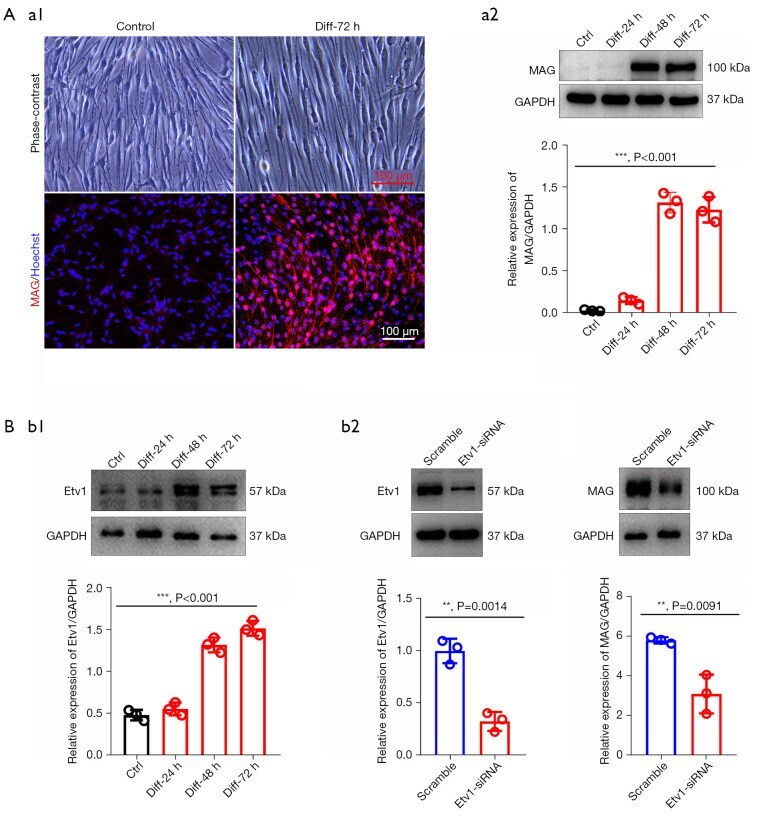
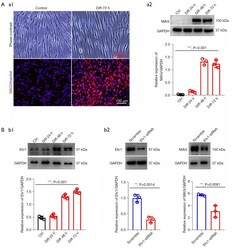
- Etv1 knockdown in Schwann cells antagonizes its differentiation. (A) (a1) Representative images of phase contrast microscopy and ICC analysis, showing that dB-cAMP can effectively induce SC differentiation. Red staining represents MAG expression and cell nuclei (blue) are marked with Hoechst staining. Scale bar =100 mum. (a2) WB comparing the expression of MAG in SCs treated with dB-cAMP or vehicle for 24, 48, and 72 hours. ***, P < 0.001; one-way ANOVA; n=3 per group. (B) (b1) Western blot comparing Etv1 levels in SCs differentiated for 24, 48, and 72 hours. ***, P < 0.001; one-way ANOVA; n=3 per group. (b2) WB showing Etv1 and MAG expression in differentiated SCs transfected with Etv1-siRNA and scrambled siRNA. The histogram shows that the knockdown of Etv1 in SCs significantly reduced the expression of MAG after the induction of differentiation. Student's t -test; **, P < 0.01 vs. scramble; n=3 per group. MAG, myelin-associated glycoprotein; GAPDH, glyceraldehyde 3-phosphate dehydrogenase; Ctrl, control; Dif, differentiation; siRNA, small interfering RNA; ICC, immunocytochemistry; SC, Schwann cell; WB, Western blotting; ANOVA, analysis of variance.