 Explore
 Validate
 Learn
Learn Western blot
 Immunocytochemistry
Antibody data
- Antibody Data
- Antigen structure
- References [0]
- Comments [0]
- Validations
- Immunocytochemistry [1]
- Immunohistochemistry [7]
Submit
Validation data
Reference
Comment
Report error
- Product number
- MA5-26999 - Provider product page

- Provider
- Invitrogen Antibodies
- Product name
- MSP Monoclonal Antibody (OTI1A10)
- Antibody type
- Monoclonal
- Antigen
- Recombinant protein fragment
- Reactivity
- Human, Mouse
- Host
- Mouse
- Isotype
- IgG
- Antibody clone number
- OTI1A10
- Vial size
- 100 μL
- Concentration
- 1 mg/mL
- Storage
- -20°C, Avoid Freeze/Thaw Cycles
No comments: Submit comment
Supportive validation
- Submitted by
- Invitrogen Antibodies (provider)
- Main image
- Experimental details
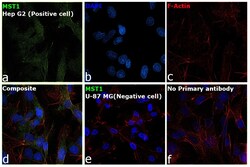
- Immunofluorescence analysis of MST1 was performed using 70% confluent log phase Hep G2 cells. The cells were fixed with 4% paraformaldehyde for 15 minutes, permeabilized with 0.1% Triton™ X-100 for 15 minutes, and blocked with 2% BSA for 15 minutes at room temperature. The cells were labeled with MST1 Monoclonal Antibody (OTI1A10) (Product # MA5-26999) at 1:100 dilution in 0.1% BSA, incubated at 4 degree celsius overnight and then labeled with Goat anti-Mouse IgG (H+L) Superclonal™ Recombinant Secondary Antibody, Alexa Fluor® 488 conjugate (Product # A28175), (1:2000 dilution), for 45 minutes at room temperature (Panel a: Green). Nuclei (Panel b: Blue) were stained with ProLong™ Diamond Antifade Mountant with DAPI (Product # P36962). F-actin (Panel c: Red) was stained with Rhodamine Phalloidin (Product # R415, 1:300 dilution). Panel d represents the merged image showing membrane and cytoplasm localization. Panel e represents U-87 MG cells showing no expression of MST1. Panel f represents control Hep G2 cells with no primary antibody to assess background. The images were captured at 60X magnification.
Supportive validation
- Submitted by
- Invitrogen Antibodies (provider)
- Main image

- Experimental details
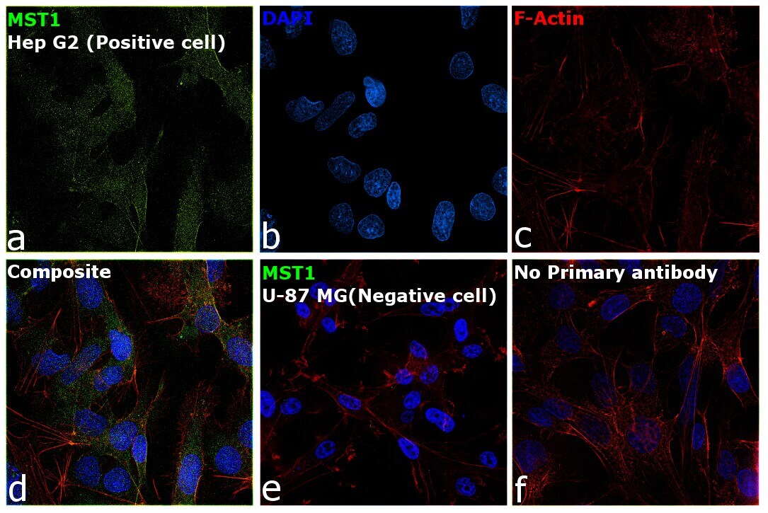
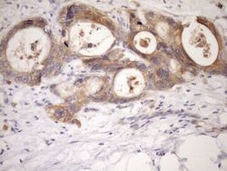
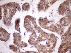
- Immunohistochemistry was performed on paraffin-embedded adenocarcinoma of human breast tissue. To expose target proteins, heat-induced epitope retrieval by Tris-EDTA, pH8.0. Following antigen retrieval, tissues were probed with a MST1 monoclonal antibody (Product # MA5-26999) at a dilution of 1:150.
- Submitted by
- Invitrogen Antibodies (provider)
- Main image

- Experimental details
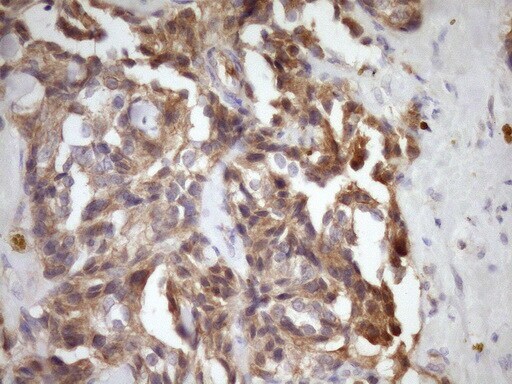
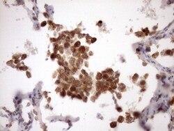
- Immunohistochemistry was performed on paraffin-embedded human colon tissue. To expose target proteins, heat-induced epitope retrieval by Tris-EDTA, pH8.0. Following antigen retrieval, tissues were probed with a MST1 monoclonal antibody (Product # MA5-26999) at a dilution of 1:150.
- Submitted by
- Invitrogen Antibodies (provider)
- Main image

- Experimental details
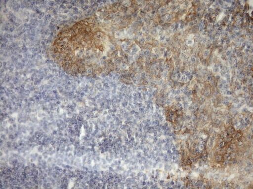
- Immunohistochemistry was performed on paraffin-embedded human tonsil tissue. To expose target proteins, heat-induced epitope retrieval by Tris-EDTA, pH8.0. Following antigen retrieval, tissues were probed with a MST1 monoclonal antibody (Product # MA5-26999) at a dilution of 1:150.
- Submitted by
- Invitrogen Antibodies (provider)
- Main image
- Experimental details
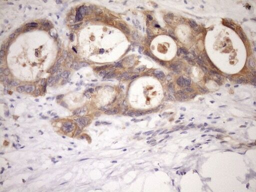
- Immunohistochemistry was performed on paraffin-embedded adenocarcinoma of human colon tissue. To expose target proteins, heat-induced epitope retrieval by Tris-EDTA, pH8.0. Following antigen retrieval, tissues were probed with a MST1 monoclonal antibody (Product # MA5-26999) at a dilution of 1:150.
- Submitted by
- Invitrogen Antibodies (provider)
- Main image
- Experimental details
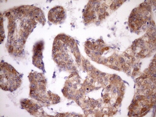
- Immunohistochemistry was performed on paraffin-embedded carcinoma of human kidney tissue. To expose target proteins, heat-induced epitope retrieval by Tris-EDTA, pH8.0. Following antigen retrieval, tissues were probed with a MST1 monoclonal antibody (Product # MA5-26999) at a dilution of 1:150.

- Submitted by
- Invitrogen Antibodies (provider)
- Main image
- Experimental details
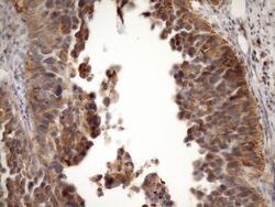
- Immunohistochemistry was performed on paraffin-embedded carcinoma of human liver tissue. To expose target proteins, heat-induced epitope retrieval by Tris-EDTA, pH8.0. Following antigen retrieval, tissues were probed with a MST1 monoclonal antibody (Product # MA5-26999) at a dilution of 1:150.
- Submitted by
- Invitrogen Antibodies (provider)
- Main image
- Experimental details
- Immunohistochemistry was performed on paraffin-embedded carcinoma of human lung tissue. To expose target proteins, heat-induced epitope retrieval by Tris-EDTA, pH8.0. Following antigen retrieval, tissues were probed with a MST1 monoclonal antibody (Product # MA5-26999) at a dilution of 1:150.
