Explore
 Validate
 Learn
Learn Western blot
Antibody data
- Antibody Data
- Antigen structure
- References [0]
- Comments [0]
- Validations
- Western blot [2]
- Immunohistochemistry [1]
- Flow cytometry [1]
Submit
Validation data
Reference
Comment
Report error
- Product number
- AP50312PU-N - Provider product page

- Provider
- Acris Antibodies GmbH
- Proper citation
- Acris Antibodies GmbH Cat#AP50312PU-N, RRID:AB_11142812
- Product name
- anti Ataxin-3 (Center)
- Antibody type
- Polyclonal
- Antigen
- KLH conjugated synthetic peptide between 267~297 amino acids from the Center region of human ATXN3
- Reactivity
- Human
- Host
- Rabbit
- Vial size
- 0.4 ml
- Concentration
- lot specific
No comments: Submit comment
Supportive validation
- Submitted by
- Acris Antibodies GmbH (provider)
- Main image
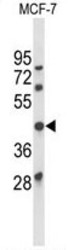
- Experimental details
- Western blot analysis of ATXN3 Antibody (Center) (Cat. #AP50312PU-N) in MCF-7 cell line lysates (35µg/lane). ATXN3 (arrow) was detected using the purified Pab.

- Submitted by
- Acris Antibodies GmbH (provider)
- Main image

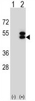
- Experimental details
- Western blot analysis of ATXN3 (arrow) using rabbit polyclonal ATXN3 Antibody (Center) (Cat. #AP50312PU-N). 293 cell lysates (2 µg/lane) either nontransfected (Lane 1) or transiently transfected (Lane 2) with the ATXN3 gene.
Supportive validation
- Submitted by
- Acris Antibodies GmbH (provider)
- Main image

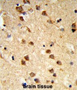
- Experimental details
- Formalin-fixed and paraffin-embedded human brain tissue reacted with ATXN3 Antibody (Center), which was peroxidase-conjugated to the secondary antibody, followed by DAB staining. This data demonstrates the use of this antibody for immunohistochemistry; clinical relevance has not been evaluated.
Supportive validation
- Submitted by
- Acris Antibodies GmbH (provider)
- Main image

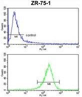
- Experimental details
- ATXN3 Antibody (Center) (Cat.#AP50312PU-N) FC analysis of ZR-75-1 cells (bottom histogram) compared to a negative control cell (top histogram). FITC-conjugated goat-anti-rabbit secondary antibodies were used for the analysis.