Explore
 Validate
 Learn
Learn Western blot
Antibody data
- Antibody Data
- Antigen structure
- References [0]
- Comments [0]
- Validations
- Western blot [3]
- Immunocytochemistry [1]
Submit
Validation data
Reference
Comment
Report error
- Product number
- MA3-082 - Provider product page

- Provider
- Invitrogen Antibodies
- Product name
- Ataxin 3 Monoclonal Antibody (2SCA-1H9)
- Antibody type
- Monoclonal
- Antigen
- Other
- Description
- MA3-082 detects Ataxin 3 from human, mouse and rat samples. MA3-082 has been successfully used in Western blot, immunofluorescence, and ELISA applications. MA3-082 has been successfully used in Western blot, immunofluorescence, and ELISA applications. The MA3-082 immunogen is the human ataxin-3 fragment from aa 112 - aa L249 as a fusion protein.
- Reactivity
- Human, Mouse, Rat
- Host
- Mouse
- Isotype
- IgG
- Antibody clone number
- 2SCA-1H9
- Vial size
- 50 µL
- Concentration
- Conc. Not Determined
- Storage
- -20° C, Avoid Freeze/Thaw Cycles
No comments: Submit comment
Supportive validation
- Submitted by
- Invitrogen Antibodies (provider)
- Main image

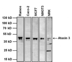
- Experimental details
- Western blot analysis of Ataxin 3 was performed by loading 20 µg of the indicated whole cell lysates and 5 µL of PageRuler Plus Prestained Protein Ladder (Product # 26619) per well onto a 4-20% Tris-Glycine polyacrylamide gel (Product # WT4202BX10). Proteins were transferred to a nitrocellulose membrane using the G2 Blotter (Product # 62288), and blocked with 5% Milk in TBST for 1 hour at room temperature. Ataxin 3 was detected at 42 kDa using a Ataxin 3 mouse monoclonal antibody (Product # MA3-082) at a dilution of 1:500 in blocking buffer for 1 hour at room temperature on a rocking platform, followed by a Goat anti-Mouse IgG (H+L) Superclonal™ Secondary Antibody, HRP conjugate (Product # A28177) at a dilution of 1:1000 for at least 30 minutes at room temperature. Chemiluminescent detection was performed using SuperSignal West Pico substrat0e (Product # 34078) and the myECL Imager (Product # 62236).

- Submitted by
- Invitrogen Antibodies (provider)
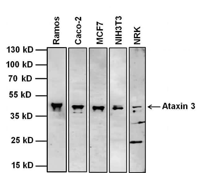
- Main image
- Experimental details
- Western blot analysis of Ataxin 3 was performed by loading 20 µg of the indicated whole cell lysates and 5 µL of PageRuler Plus Prestained Protein Ladder (Product # 26619) per well onto a 4-20% Tris-Glycine polyacrylamide gel (Product # WT4202BX10). Proteins were transferred to a nitrocellulose membrane using the G2 Blotter (Product # 62288), and blocked with 5% Milk in TBST for 1 hour at room temperature. Ataxin 3 was detected at 42 kDa using a Ataxin 3 mouse monoclonal antibody (Product # MA3-082) at a dilution of 1:500 in blocking buffer for 1 hour at room temperature on a rocking platform, followed by a Goat anti-Mouse IgG (H+L) Superclonal™ Secondary Antibody, HRP conjugate (Product # A28177) at a dilution of 1:1000 for at least 30 minutes at room temperature. Chemiluminescent detection was performed using SuperSignal West Pico substrat0e (Product # 34078) and the myECL Imager (Product # 62236).
- Submitted by
- Invitrogen Antibodies (provider)
- Main image

- Experimental details
- Western blot analysis of Ataxin 3 was performed by loading 20 µg of HEK293T wildtype (Lane 1), HEK293T Ataxin 3 knockout (Lane 2) whole cell extracts. The blot was probed with Anti-Ataxin 3 Monoclonal Antibody (Product # MA3-082) (1:1000 dilution) and Goat anti-Mouse IgG (H+L), Superclonal™ Recombinant Secondary Antibody, HRP (Product # A28177) (1:4000 dilution). Loss of signal upon CRISPR mediated knockout (KO) confirms that antibody is specific to Ataxin 3.

Supportive validation
- Submitted by
- Invitrogen Antibodies (provider)
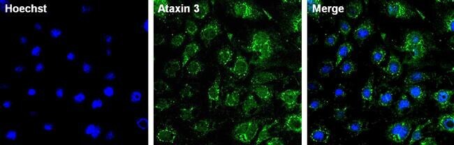
- Main image
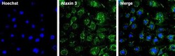
- Experimental details
- Immunofluorescent analysis of Ataxin 3 (green) in Caco-2 cells. The cells were fixed with 4% paraformaldehyde for 15 minutes, permeabilized with 0.1% Triton X-100 in PBS for 15 minutes, and blocked with 3% BSA in PBS (Product # 37525) for 30 minutes at room temperature. Cells were stained with a Ataxin 3 mouse monoclonal antibody (Product # MA3-082) at a dilution of 1:100 in staining buffer for 1 hour at room temperature, and then incubated with a Goat anti-Mouse IgG Superclonal™ Secondary Antibody, Alexa Fluor 488 conjugate (Product # A28175) at a dilution of 1:1000 for 1 hour at room temperature. Nuclei (blue) were counterstained with Hoechst 33342 dye (Product # 62249). Images were taken on a Thermo Scientific ToxInsight Instrument at 20X magnification.