Explore
 Validate
 Learn
Learn Western blot
Antibody data
- Antibody Data
- Antigen structure
- References [1]
- Comments [0]
- Validations
- Western blot [3]
- Flow cytometry [1]
- Other assay [1]
Submit
Validation data
Reference
Comment
Report error
- Product number
- PA1-514 - Provider product page

- Provider
- Invitrogen Antibodies
- Product name
- AIP Polyclonal Antibody
- Antibody type
- Polyclonal
- Antigen
- Recombinant full-length protein
- Description
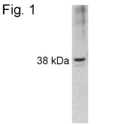
- PA1-514 detects aryl hydrocarbon receptor interacting protein (AIP) from mouse samples. PA1-514 has been successfully used in Western blot procedures. By Western blot, this antibody detects an ~38 kDa protein representing AIP from Hepa 1 SV40 cell lysate, as well as recombinant mouse AIP. PA1-514 immunogen is full length, bacterially expressed mouse recombinant AIP.
- Reactivity
- Human, Mouse
- Host
- Rabbit
- Isotype
- IgG
- Vial size
- 100 μg
- Concentration
- 1 mg/mL
- Storage
- -20°C, Avoid Freeze/Thaw Cycles
Submitted references The cholinergic and non-cholinergic effects of organophosphates and oximes in cultured human myoblasts.
Katalinić M, Miš K, Pirkmajer S, Grubič Z, Kovarik Z, Marš T
Chemico-biological interactions 2013 Mar 25;203(1):144-8
Chemico-biological interactions 2013 Mar 25;203(1):144-8
No comments: Submit comment
Supportive validation
- Submitted by
- Invitrogen Antibodies (provider)
- Main image
- Experimental details
- Western blot of recombinant mouse AIP using Product # PA1-514.

- Submitted by
- Invitrogen Antibodies (provider)
- Main image
- Experimental details
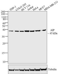
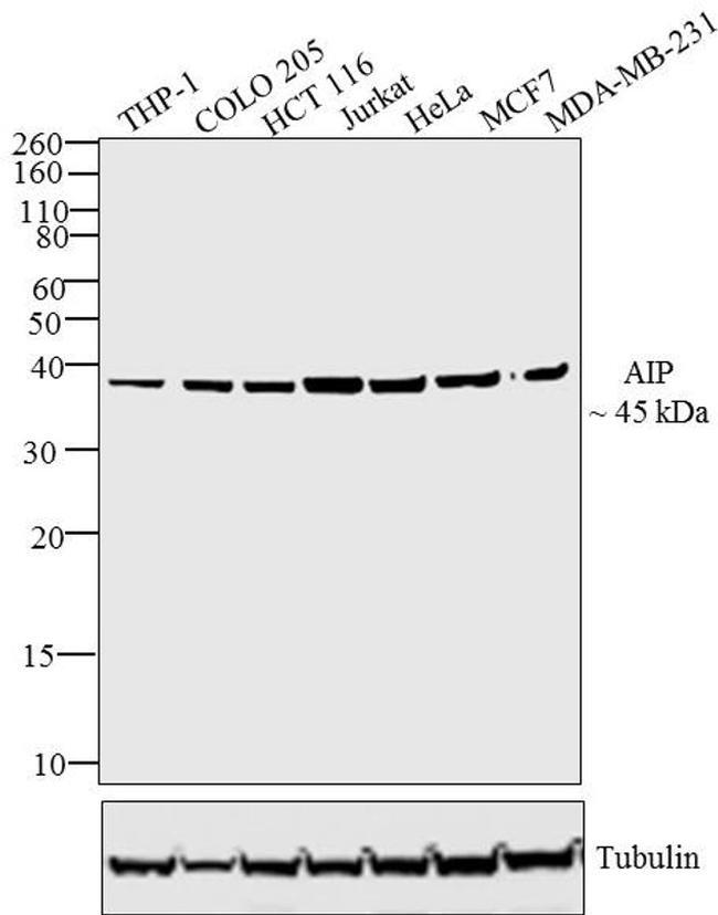
- Western blot analysis was performed on whole cell extracts (30 µg) of THP-1 (Lane 1), COLO 205 (Lane 2), HCT 116 (Lane 3), Jurkat (Lane 4), HeLa (Lane 5), MCF7 (Lane 6) and MDA-MB-231 (Lane 7).The blots were probed with Anti-AIP Rabbit Polyclonal Antibody (Product # PA1-514, 2 µg/mL) and detected by chemiluminescence using Goat anti-Rabbit IgG (Heavy Chain) Superclonal™ Secondary Antibody, HRP conjugate (Product # A27036, 0.4 µg/mL, 1:2500 dilution). A ~45 kDa band corresponding to AIP was observed across cell lines tested. Known quantity of protein samples were electrophoresed using Novex® NuPAGE®12 % Bis-Tris gel (Product # NP0342BOX), XCell SureLock™ Electrophoresis System (Product # EI0002) and Novex® Sharp Pre-Stained Protein Standard (Product # LC5800). Resolved proteins were then transferred onto a nitrocellulose membrane iBlot® 2 Dry Blotting System (Product # IB21001). The membrane was probed with the relevant primary and secondary Antibody using iBind™ Flex Western Starter Kit (Product # SLF2000S). Chemiluminescent detection was performed using Pierce™ ECL Western Blotting Substrate (Product # 32106).
- Submitted by
- Invitrogen Antibodies (provider)
- Main image

- Experimental details
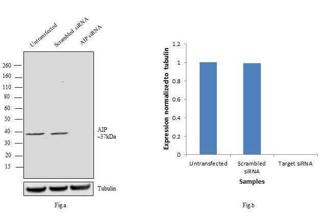
- Knockdown of AIP was achieved by transfecting HeLa cells with AIP specific siRNAs (Silencer® select Product # s17251, s531246). Western blot analysis (Fig a) was performed using whole cell extracts from the AIP knock down cells (lane 3), non-specific scrambled siRNA transfected cells (lane 2) and untransfected cells (lane 1). The blots were probed with Anti-AIP Rabbit polyclonal Antibody (Product # PA1-514, 2µg/mL) and Goat anti-Rabbit IgG (Heavy Chain) Superclonal™ Secondary Antibody, HRP conjugate (Product # A27036, 0.4 µg/mL, 1:4000 dilution). Densitometric analysis of this western blot is shown in histogram(Fig b). Loss of signal upon siRNA mediated knock down confirms that antibody is specific to AIP.
Supportive validation
- Submitted by
- Invitrogen Antibodies (provider)
- Main image

- Experimental details
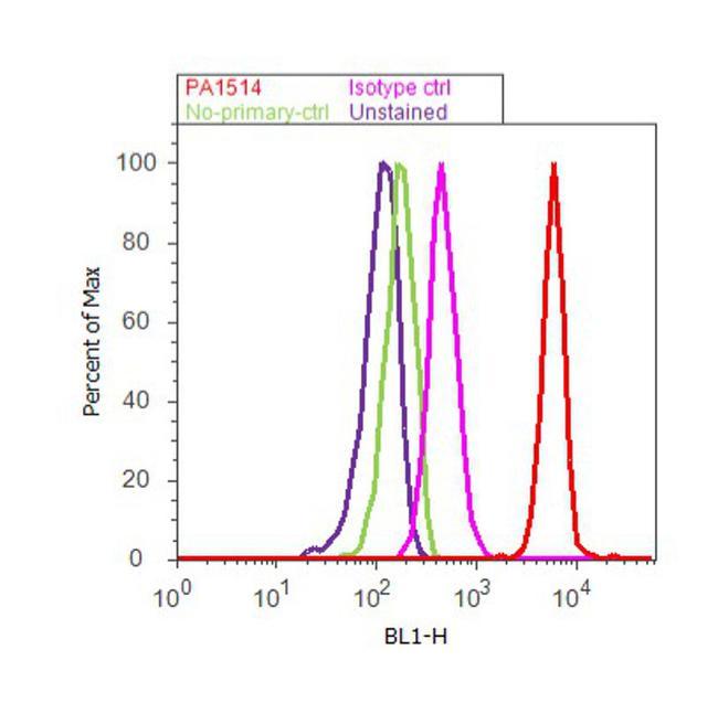
- Flow cytometry analysis of AIP was done on A549 cells. Cells were fixed with 70% ethanol for 10 minutes, permeabilized with 0.25% Triton™ X-100 for 20 minutes, and blocked with 5% BSA for 30 minutes at room temperature. Cells were labeled with AIP Rabbit Polyclonal Antibody (PA1-514, red histogram) or with rabbit isotype control (pink histogram) at 3-5 ug/million cells in 2.5% BSA. After incubation at room temperature for 2 hours, the cells were labeled with Alexa Fluor® 488 Goat Anti-Rabbit Secondary Antibody (A11008) at a dilution of 1:400 for 30 minutes at room temperature. The representative 10, 000 cells were acquired and analyzed for each sample using an Attune® Acoustic Focusing Cytometer. The purple histogram represents unstained control cells and the green histogram represents no-primary-antibody control.
Supportive validation
- Submitted by
- Invitrogen Antibodies (provider)
- Main image

- Experimental details
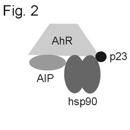
- Diagram of the AIP-hsp90-AhR complex