 Explore
 Validate
 Learn
Learn Western blot
 Immunoprecipitation
 Immunohistochemistry
Antibody data
- Antibody Data
- Antigen structure
- References [1]
- Comments [0]
- Validations
- Immunohistochemistry [1]
- Flow cytometry [2]
Submit
Validation data
Reference
Comment
Report error
- Product number
- MA3-16515 - Provider product page

- Provider
- Invitrogen Antibodies
- Product name
- AIP Monoclonal Antibody (35-2)
- Antibody type
- Monoclonal
- Antigen
- Other
- Description
- This antibody is specific for the FKBP domain. Suggested positive control: antigen standard for AIP (transient overexpression lysate).
- Reactivity
- Human, Mouse, Rat
- Host
- Mouse
- Isotype
- IgG
- Antibody clone number
- 35-2
- Vial size
- 100 μL
- Concentration
- Conc. Not Determined
- Storage
- -20°C, Avoid Freeze/Thaw Cycles
Submitted references Mechanistic insights into cancer cell killing through interaction of phosphodiesterase 3A and schlafen family member 12.
Wu X, Schnitzler GR, Gao GF, Diamond B, Baker AR, Kaplan B, Williamson K, Westlake L, Lorrey S, Lewis TA, Garvie CW, Lange M, Hayat S, Seidel H, Doench J, Cherniack AD, Kopitz C, Meyerson M, Greulich H
The Journal of biological chemistry 2020 Mar 13;295(11):3431-3446
The Journal of biological chemistry 2020 Mar 13;295(11):3431-3446
No comments: Submit comment
Supportive validation
- Submitted by
- Invitrogen Antibodies (provider)
- Main image
- Experimental details
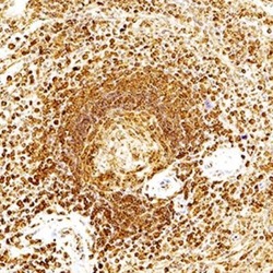
- Immunohistochemical analysis of AIP in immersion fixed paraffin-embedded sections of human spleen. Samples were incubated in AIP monoclonal antibody (Product # MA3-16515) using a dilution of 1:300 for 1 hour at room temperature followed by the Anti-Mouse IgG VisUCyte™ HRP Polymer Antibody. Tissue was stained using DAB (brown) and counterstained with hematoxylin (blue). Specific staining was localized to the cytoplasm in splenocytes.

Supportive validation
- Submitted by
- Invitrogen Antibodies (provider)
- Main image
- Experimental details
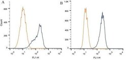
- Flow cytometry of AIP in 1 x 10^6 CHO (A) and MCF-7 (B) cells. Samples were incubated in AIP monoclonal antibody (Product # MA3-16515) using a dilution of 1 µg/1x10^6 cells. Antibody (dark blue). Isotype control shown in orange.
- Submitted by
- Invitrogen Antibodies (provider)
- Main image

- Experimental details
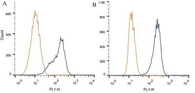
- Flow cytometry of AIP in 1 x 10^6 CHO (A) and MCF-7 (B) cells. Samples were incubated in AIP monoclonal antibody (Product # MA3-16515) using a dilution of 1 µg/1x10^6 cells. Antibody (dark blue). Isotype control shown in orange.
