Explore
 Validate
 Learn
Learn Immunohistochemistry
 Flow cytometry
Antibody data
- Antibody Data
- Antigen structure
- References [6]
- Comments [0]
- Validations
- Flow cytometry [1]
- Other assay [1]
Submit
Validation data
Reference
Comment
Report error
- Product number
- 14-0119-82 - Provider product page

- Provider
- Invitrogen Antibodies
- Product name
- CD11a (LFA-1alpha) Monoclonal Antibody (HI111), eBioscience™
- Antibody type
- Monoclonal
- Antigen
- Other
- Description
- Description: The HI111 monoclonal antibody reacts with human CD11a, the 180 kDa integrin alphaL, also known as the lymphocyte function associated antigen-1 (LFA-1) alpha chain. LFA-1, formed by non-covalent association of CD11a with CD18 (integrin beta2), serves as an important adhesion molecule involved in lymphocyte and granulocyte function. CD54 (ICAM-1), CD102 (ICAM-2), and CD50 (ICAM-3) are ligands for LFA-1. CD11a is expressed by all leukocytes. Applications Reported: This HI111 antibody has been reported for use in flow cytometric analysis, and immunohistology staining of frozen tissue sections. It has also been reported in blocking of CD11a function in mixed lymphocyte reactions. (Please use Functional Grade purified HI111, Product # 16-0119, in functional assays). Applications Tested: The HI111 antibody has been tested by flow cytometric analysis of normal human peripheral blood cells. This can be used at less than or equal to 1 µg per test. A test is defined as the amount (µg) of antibody that will stain a cell sample in a final volume of 100 µL. Cell number should be determined empirically but can range from 10^5 to 10^8 cells/test. It is recommended that the antibody be carefully titrated for optimal performance in the assay of interest. Purity: Greater than 90%, as determined by SDS-PAGE. Aggregation: Less than 10%, as determined by HPLC. Filtration: 0.2 µm post-manufacturing filtered.
- Reactivity
- Human
- Host
- Mouse
- Isotype
- IgG
- Antibody clone number
- HI111
- Vial size
- 100 μg
- Concentration
- 0.5 mg/mL
- Storage
- 4°C
Submitted references Failure to upregulate cell surface PD-1 is associated with dysregulated stimulation of T cells by TGN1412-like CD28 superagonist.
Image correlation microscopy for uniform illumination.
Comparison of gene expression profiles between human and mouse monocyte subsets.
Expression of endothelia and lymphocyte adhesion molecules in bronchus-associated lymphoid tissue (BALT) in adult human lung.
Systemic hypoxia enhances bactericidal activities of human polymorphonuclear leuocytes.
Activation of epithelial and myoepithelial cells in the salivary glands of patients with Sjögren's syndrome: high expression of intercellular adhesion molecule-1 (ICAM.1) in biopsy specimens and cultured cells.
Thaventhiran T, Alhumeed N, Yeang HX, Sethu S, Downey JS, Alghanem AF, Olayanju A, Smith EL, Cross MJ, Webb SD, Williams DP, Bristow A, Ball C, Stebbings R, Sathish JG
mAbs 2014;6(5):1290-9
mAbs 2014;6(5):1290-9
Image correlation microscopy for uniform illumination.
Gaborski TR, Sealander MN, Ehrenberg M, Waugh RE, McGrath JL
Journal of microscopy 2010 Jan;237(1):39-50
Journal of microscopy 2010 Jan;237(1):39-50
Comparison of gene expression profiles between human and mouse monocyte subsets.
Ingersoll MA, Spanbroek R, Lottaz C, Gautier EL, Frankenberger M, Hoffmann R, Lang R, Haniffa M, Collin M, Tacke F, Habenicht AJ, Ziegler-Heitbrock L, Randolph GJ
Blood 2010 Jan 21;115(3):e10-9
Blood 2010 Jan 21;115(3):e10-9
Expression of endothelia and lymphocyte adhesion molecules in bronchus-associated lymphoid tissue (BALT) in adult human lung.
Kawamata N, Xu B, Nishijima H, Aoyama K, Kusumoto M, Takeuchi T, Tei C, Michie SA, Matsuyama T
Respiratory research 2009 Oct 22;10(1):97
Respiratory research 2009 Oct 22;10(1):97
Systemic hypoxia enhances bactericidal activities of human polymorphonuclear leuocytes.
Wang JS, Liu HC
Clinical science (London, England : 1979) 2009 May 1;116(11):805-17
Clinical science (London, England : 1979) 2009 May 1;116(11):805-17
Activation of epithelial and myoepithelial cells in the salivary glands of patients with Sjögren's syndrome: high expression of intercellular adhesion molecule-1 (ICAM.1) in biopsy specimens and cultured cells.
Kapsogeorgou EK, Dimitriou ID, Abu-Helu RF, Moutsopoulos HM, Manoussakis MN
Clinical and experimental immunology 2001 Apr;124(1):126-33
Clinical and experimental immunology 2001 Apr;124(1):126-33
No comments: Submit comment
Supportive validation
- Submitted by
- Invitrogen Antibodies (provider)
- Main image
- Experimental details
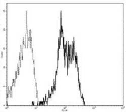
- Staining of normal human peripheral blood cells with Anti-Human CD11a FITC.

Supportive validation
- Submitted by
- Invitrogen Antibodies (provider)
- Main image
- Experimental details
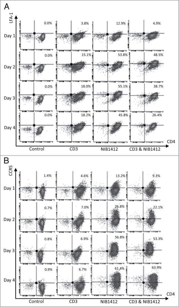
- Figure 2. Enhanced cell surface expression of LFA-1 and CCR5 on CD28SA-activated CD4 + effector memory T cells. Human CD4 + T EMs were stimulated for 1 to 4 d with plate-bound anti-CD3 mAb (CD3, 5 mug/ml); NIB1412 (NIB1412, 10 mug/ml); anti-CD3 mAb and NIB1412 (CD3&NIB1412); control category included cells without any treatment (Control). Cells were harvested at indicated time points and stained with fluorochrome-conjugated anti-CD4 and anti-LFA ( A ) or anti-CD4 and anti-CCR5 ( B ) antibodies followed by flow cytometric analysis. ( A ) Population of CD4 + LFA + cells are shown in the upper right quadrant as percentages of total T cells. The cells are shown as percentages of total T cells in the upper right quadrant. ( B ) Population of CD4 + CCR5 + cells are shown in the upper right quadrant as percentages of total T cells. Results are representative of four independent experiments.