Explore
 Validate
 Learn
LearnAPZ-045-200UL
antibody from Invitrogen Antibodies
Targeting: GIPC1
C19orf3, GIPC, GLUT1CBP, Hs.6454, NIP, RGS19IP1, SEMCAP, SYNECTIN, TIP-2
 Western blot
Antibody data
- Antibody Data
- Antigen structure
- References [0]
- Comments [0]
- Validations
- Western blot [3]
- Immunohistochemistry [1]
Submit
Validation data
Reference
Comment
Report error
- Product number
- APZ-045-200UL - Provider product page

- Provider
- Invitrogen Antibodies
- Product name
- GIPC1 Polyclonal Antibody
- Antibody type
- Polyclonal
- Antigen
- Other
- Reactivity
- Human, Mouse, Rat
- Host
- Rabbit
- Isotype
- IgG
- Vial size
- 200 µL
- Concentration
- 0.8 mg/mL
- Storage
- -20° C, Avoid Freeze/Thaw Cycles
No comments: Submit comment
Supportive validation
- Submitted by
- Invitrogen Antibodies (provider)
- Main image

- Experimental details
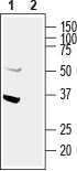
- Western blot analysis of human U87-MG glioblastoma cell line lysate: - 1. Anti-GIPC1 Antibody (#APZ-045), (1:400). 2. Anti-GIPC1 Antibody , preincubated with GIPC1 Blocking Peptide (#BLP-PZ045).
- Submitted by
- Invitrogen Antibodies (provider)
- Main image
- Experimental details
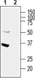
- Western blot analysis of human U87-MG glioblastoma cell line lysate: - 1. Anti-GIPC1 Antibody (#APZ-045), (1:400). 2. Anti-GIPC1 Antibody , preincubated with GIPC1 Blocking Peptide (#BLP-PZ045).

- Submitted by
- Invitrogen Antibodies (provider)
- Main image
- Experimental details
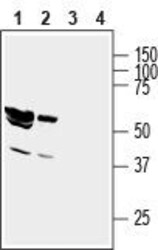
- Western blot analysis of rat brain lysate (lanes 1 and 3) and mouse brain membranes (lanes 2 and 4): - 1,2. Anti-GIPC1 Antibody (#APZ-045), (1:400).3,4. Anti-GIPC1 Antibody , preincubated with GIPC1 Blocking Peptide (#BLP-PZ045).

Supportive validation
- Submitted by
- Invitrogen Antibodies (provider)
- Main image
- Experimental details
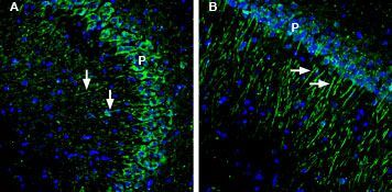
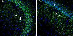
- Expression of GIPC1 in mouse and rat hippocampus - Immunohistochemical staining of perfusion-fixed frozen mouse and rat brain sections with Anti-GIPC1 Antibody (#APZ-045), (1:200), followed by goat- Anti-rabbit-AlexaFluor-488. A. In mouse brain GIPC1 staining (green) appears in neurons of the pyramidal layer (P) and their apical dendrites (arrows). B. In rat brain similar staining is observed. Cell nuclei are stained with DAPI (blue).