Explore
 Validate
 Learn
Learn Western blot
 Immunoprecipitation
 Other assay
Antibody data
- Antibody Data
- Antigen structure
- References [4]
- Comments [0]
- Validations
- Other assay [2]
Submit
Validation data
Reference
Comment
Report error
- Product number
- 36-5800 - Provider product page

- Provider
- Invitrogen Antibodies
- Product name
- WNT1 Polyclonal Antibody
- Antibody type
- Polyclonal
- Antigen
- Synthetic peptide
- Reactivity
- Human, Mouse
- Host
- Rabbit
- Isotype
- IgG
- Vial size
- 100 μg
- Concentration
- 0.25 mg/mL
- Storage
- -20°C
Submitted references The loss of Krüppel-like factor 15 in Foxd1(+) stromal cells exacerbates kidney fibrosis.
miR-148a is Associated with Obesity and Modulates Adipocyte Differentiation of Mesenchymal Stem Cells through Wnt Signaling.
Overexpression of Wnt-1 in thyrocytes enhances cellular growth but suppresses transcription of the thyroperoxidase gene via different signaling mechanisms.
Axin is a scaffold protein in TGF-beta signaling that promotes degradation of Smad7 by Arkadia.
Gu X, Mallipattu SK, Guo Y, Revelo MP, Pace J, Miller T, Gao X, Jain MK, Bialkowska AB, Yang VW, He JC, Mei C
Kidney international 2017 Nov;92(5):1178-1193
Kidney international 2017 Nov;92(5):1178-1193
miR-148a is Associated with Obesity and Modulates Adipocyte Differentiation of Mesenchymal Stem Cells through Wnt Signaling.
Shi C, Zhang M, Tong M, Yang L, Pang L, Chen L, Xu G, Chi X, Hong Q, Ni Y, Ji C, Guo X
Scientific reports 2015 May 22;5:9930
Scientific reports 2015 May 22;5:9930
Overexpression of Wnt-1 in thyrocytes enhances cellular growth but suppresses transcription of the thyroperoxidase gene via different signaling mechanisms.
Kim WB, Lewis CJ, McCall KD, Malgor R, Kohn AD, Moon RT, Kohn LD
The Journal of endocrinology 2007 Apr;193(1):93-106
The Journal of endocrinology 2007 Apr;193(1):93-106
Axin is a scaffold protein in TGF-beta signaling that promotes degradation of Smad7 by Arkadia.
Liu W, Rui H, Wang J, Lin S, He Y, Chen M, Li Q, Ye Z, Zhang S, Chan SC, Chen YG, Han J, Lin SC
The EMBO journal 2006 Apr 19;25(8):1646-58
The EMBO journal 2006 Apr 19;25(8):1646-58
No comments: Submit comment
Supportive validation
- Submitted by
- Invitrogen Antibodies (provider)
- Main image

- Experimental details
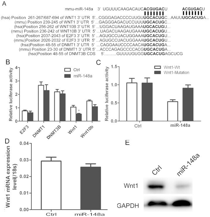
- Figure 4 Identification of miR-148a-binding sequence in target genes. (A) Predicted interaction between miR-148a and its putative binding sites inthe 3' UTR or CDS of target gene. Luciferase activity of HEK293cells cotransfected with reporter vector containing either wild-type (B) ormutant Wnt1 3'-UTR and miR-148a or control (C). (D) qRT-PCRanalysis of Wnt in hMSCs-Ad after stable infection with miR-148a or controllentivirus. (E) Western blot analysis of Wnt1 in hMSCs-Ad lysates afterstable infection with miR-148a or control lentivirus. GAPDH blot served asloading control. ** P < 0.01. Results are mean+- SEM of triplicate measurements ( n = 4).
- Submitted by
- Invitrogen Antibodies (provider)
- Main image
- Experimental details
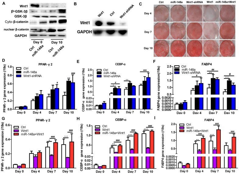
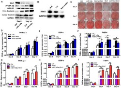
- Figure 5 miR-148a regulates adipogenesis in hMSCs-Ad via Wnt. (A) Wnt signal pathway protein level was detected by Western blot. (B)Western blot analysis of Wnt1 in hMSCs-Ad lysates after stable infectionwith Wnt1-shRNA or Wnt1 construct or control lentivirus. GAPDH blot servedas loading control. (C) Oil red O staining indicated the effects of miR-148aon hMSCs-Ad adipogenic differentiation at Days 4, 7, and 10. Transcriptionlevels of adipogenic marker genes, PPARgamma2 (D, G),C/EBP-alpha (E, H), and FABP4 (F, I), were detected by qRT-PCR indifferent groups at Days 0, 4, 7, and 10 during adipogenesis. * P < 0.05, ** P < 0.01, *** P