Explore
 Validate
 Learn
Learn Western blot
 ELISA
ELISA Immunocytochemistry
Antibody data
- Antibody Data
- Antigen structure
- References [2]
- Comments [0]
- Validations
- Immunocytochemistry [2]
- Immunohistochemistry [1]
- Flow cytometry [2]
- Other assay [1]
Submit
Validation data
Reference
Comment
Report error
- Product number
- MA5-15544 - Provider product page

- Provider
- Invitrogen Antibodies
- Product name
- WNT1 Monoclonal Antibody (10C8)
- Antibody type
- Monoclonal
- Antigen
- Purifed from natural sources
- Description
- MA5-15544 targets WNT1 in indirect ELISA, FACS, IF, IHC, and WB applications and shows reactivity with Human and mouse samples. The MA5-15544 immunogen is purified recombinant fragment of WNT1 expressed in E. Coli. MA5-15544 detects WNT1 which has a predicted molecular weight of approximately 41kDa.
- Reactivity
- Human, Mouse
- Host
- Mouse
- Isotype
- IgG
- Antibody clone number
- 10C8
- Vial size
- 100 μL
- Concentration
- Conc. Not Determined
- Storage
- Store at 4°C short term. For long term storage, store at -20°C, avoiding freeze/thaw cycles.
Submitted references E74-like factor 3 suppresses microRNA-485-5p transcription to trigger growth and metastasis of ovarian cancer cells with the involvement of CLDN4/Wnt/β-catenin axis.
Relaxin reverses maladaptive remodeling of the aged heart through Wnt-signaling.
Kuang L, Li L
Saudi journal of biological sciences 2021 Aug;28(8):4137-4146
Saudi journal of biological sciences 2021 Aug;28(8):4137-4146
Relaxin reverses maladaptive remodeling of the aged heart through Wnt-signaling.
Martin B, Gabris B, Barakat AF, Henry BL, Giannini M, Reddy RP, Wang X, Romero G, Salama G
Scientific reports 2019 Dec 6;9(1):18545
Scientific reports 2019 Dec 6;9(1):18545
No comments: Submit comment
Supportive validation
- Submitted by
- Invitrogen Antibodies (provider)
- Main image
- Experimental details
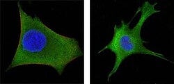
- Immunofluorescence analysis of HeLa (left) and 3T3-L1 (right) cells using WNT1 monoclonal antibody (Product # MA5-15544) (Green). Blue: DRAQ5 fluorescent DNA dye. Red: Actin filaments have been labeled with DY-554 phalloidin.

- Submitted by
- Invitrogen Antibodies (provider)
- Main image

- Experimental details
- Immunofluorescence analysis of HeLa (left) and 3T3-L1 (right) cells using WNT1 monoclonal antibody (Product # MA5-15544) (Green). Blue: DRAQ5 fluorescent DNA dye. Red: Actin filaments have been labeled with DY-554 phalloidin.

Supportive validation
- Submitted by
- Invitrogen Antibodies (provider)
- Main image
- Experimental details
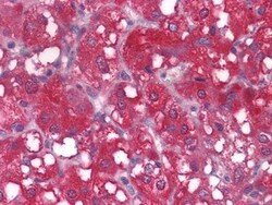
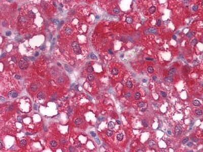
- Immunohistochemical analysis of paraffin-embedded human adrenal tissues using WNT1 monoclonal antibody (Product # MA5-15544).
Supportive validation
- Submitted by
- Invitrogen Antibodies (provider)
- Main image
- Experimental details
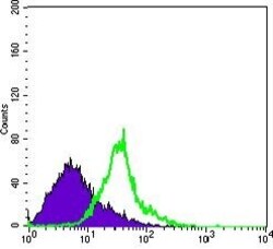
- Flow cytometric analysis of HeLa cells using WNT1 monoclonal antibody (Product # MA5-15544) (green) and negative control (purple).

- Submitted by
- Invitrogen Antibodies (provider)
- Main image
- Experimental details
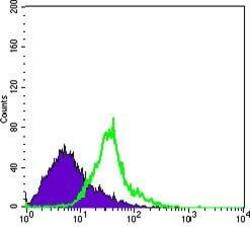
- Flow cytometric analysis of HeLa cells using WNT1 monoclonal antibody (Product # MA5-15544) (green) and negative control (purple).

Supportive validation
- Submitted by
- Invitrogen Antibodies (provider)
- Main image
- Experimental details
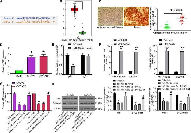
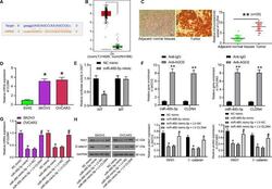
- Fig. 6 miR-485-5p targets CLDN4 to regulate the Wnt/beta-catenin signaling. A, binding site between miR-485-5p and CLDN4 predicted on Starbase ( http://starbase.sysu.edu.cn/ ); B, miR-485-5p expression profile in OC predicted on GEPIA ( http://gepia.cancer-pku.cn/ ); C, protein expression of CLDN4 in tumor tissues and the paired normal tissues determined by immunohistochemical staining (paired t test,** p < 0.01); D, CLDN4 expression in OC cells and SV40 cells measured by RT-qPCR; E-F, binding relationship between miR-485-5p and CLDN4 validated through a dual luciferase reporter gene assay (E) and an RIP assay (F) (two-way ANOVA,* p < 0.05); G, CLDN4 expression in OC cells after miR-485-5p mimic and LV-CLDN4 transfection determined by RT-qPCR (one-way ANOVA, * p < 0.05 compared to NC mimic, # p < 0.05 compared to miR-485-5p mimic + LV-NC); H, protein levels of Wnt1 and beta-catenin in OC cell lines after miR-485-5p mimic and LV-CLDN4 transfection evaluated using western blot analysis (two-way ANOVA,* p < 0.05 compared to NC mimic, # p < 0.05 compared to miR-485-5p mimic + LV-NC). Data were exhibited as mean +- SD from three independent experiments.