Explore
 Validate
 Learn
Learn Western blot
 Immunocytochemistry
Antibody data
- Antibody Data
- Antigen structure
- References [2]
- Comments [0]
- Validations
- Immunocytochemistry [3]
- Other assay [1]
Submit
Validation data
Reference
Comment
Report error
- Product number
- MA5-23522 - Provider product page

- Provider
- Invitrogen Antibodies
- Product name
- BICD2 Monoclonal Antibody (3I3)
- Antibody type
- Monoclonal
- Antigen
- Recombinant full-length protein
- Description
- Recommended positive controls: 293T, A431, HeLa, and HepG2 cells. Predicted reactivity: Mouse (100%), Rat (100%), Rhesus Monkey (100%), Bovine (100%). Store product as a concentrated solution. Centrifuge briefly prior to opening the vial.
- Reactivity
- Human, Mouse, Rat
- Host
- Mouse
- Isotype
- IgG
- Antibody clone number
- 3I3
- Vial size
- 100 μL
- Concentration
- 1 mg/mL
- Storage
- Store at 4°C short term. For long term storage, store at -20°C, avoiding freeze/thaw cycles.
Submitted references Cytoplasmic control of intranuclear polarity by human cytomegalovirus.
MiR-4674 regulates angiogenesis in tissue injury by targeting p38K signaling in endothelial cells.
Procter DJ, Furey C, Garza-Gongora AG, Kosak ST, Walsh D
Nature 2020 Nov;587(7832):109-114
Nature 2020 Nov;587(7832):109-114
MiR-4674 regulates angiogenesis in tissue injury by targeting p38K signaling in endothelial cells.
Icli B, Li H, Pérez-Cremades D, Wu W, Ozdemir D, Haemmig S, Guimaraes RB, Manica A, Marchini JF, Orgill DP, Feinberg MW
American journal of physiology. Cell physiology 2020 Mar 1;318(3):C524-C535
American journal of physiology. Cell physiology 2020 Mar 1;318(3):C524-C535
No comments: Submit comment
Supportive validation
- Submitted by
- Invitrogen Antibodies (provider)
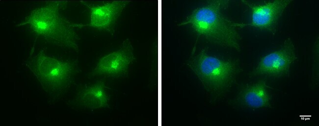
- Main image
- Experimental details
- Immunocytochemistry-Immunofluorescence analysis of BICD2 was performed in HeLa cells fixed in 4% paraformaldehyde for 10 min. Green: BICD2 Monoclonal Antibody (3I3) (Product # MA5-23522) diluted at 1:100. Blue: Hoechst 33342 staining. Scale bar = 10 µm.
- Submitted by
- Invitrogen Antibodies (provider)
- Main image

- Experimental details
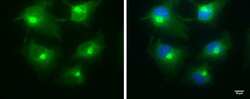
- Immunocytochemistry-Immunofluorescence analysis of BICD2 was performed in HeLa cells fixed in 4% paraformaldehyde for 10 min. Green: BICD2 Monoclonal Antibody (3I3) (Product # MA5-23522) diluted at 1:100. Blue: Hoechst 33342 staining. Scale bar = 10 µm.

- Submitted by
- Invitrogen Antibodies (provider)
- Main image
- Experimental details
- Immunocytochemistry-Immunofluorescence analysis of BICD2 was performed in HeLa cells fixed in 4% paraformaldehyde for 10 min. Green: BICD2 Monoclonal Antibody (3I3) (Product # MA5-23522) diluted at 1:100. Blue: Hoechst 33342 staining. Scale bar = 10 µm.
Supportive validation
- Submitted by
- Invitrogen Antibodies (provider)
- Main image
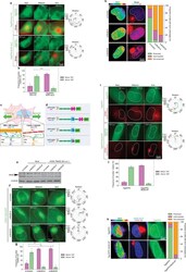
- Experimental details
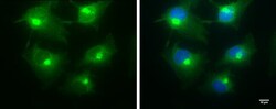
- Extended Data Fig. 3: SUN1:Nesprin-2G and the dynein adaptor BICD2 mediate nuclear rotation during HCMV infection. a-b, Expression of a SUN1 mutant that does not engage Nesprin-2G impairs nuclear rotation in HCMV-infected cells. a, Representative stills from time lapse recordings of NHDFs expressing Tag-GFP2 forms of SUN1 Full Length (FL) or SUN1 lacking the lumenal domain (SUN1DeltaLu) that mediates interactions with Nesprin-2G, infected with HCMV-UL99mCherry. Rotation traces from this imaging are shown to the right. Analyses focused on cells expressing intermediate levels of SUN1-GFP constructs as high levels of expression can result in retention of Nesprin-2G in the endoplasmic reticulum (ER). b, Quantification of rotation frequencies above or below 180deg; bars represent mean +- SEM, statistics use two-tailed student's t-test, n = 138 cells total from 3 independent biological replicates, ***pLEAA kinesin-binding mutant. e, Western blot analysis of BICD2 expressio