Explore
 Validate
 Learn
LearnHPA029424
antibody from Atlas Antibodies
Targeting: ATAD2
ANCCA, CT137, DKFZp667N1320, MGC29843, MGC5254, PRO2000
 Western blot
 Immunohistochemistry
Antibody data
- Antibody Data
- Antigen structure
- References [4]
- Comments [0]
- Validations
- Western blot [1]
- Immunocytochemistry [2]
- Immunohistochemistry [1]
Submit
Validation data
Reference
Comment
Report error
- Product number
- HPA029424 - Provider product page

- Provider
- Atlas Antibodies
- Proper citation
- Atlas Antibodies Cat#HPA029424, RRID:AB_10611680
- Product name
- Anti-ATAD2
- Antibody type
- Polyclonal
- Description
- Polyclonal Antibody against Human ATAD2, Gene description: ATPase family, AAA domain containing 2, Alternative Gene Names: CT137, DKFZp667N1320, MGC29843, MGC5254, PRO2000, Validated applications: ICC, IHC, WB, Uniprot ID: Q6PL18, Storage: Store at +4°C for short term storage. Long time storage is recommended at -20°C.
- Reactivity
- Human
- Host
- Rabbit
- Conjugate
- Unconjugated
- Isotype
- IgG
- Vial size
- 100 µl
- Concentration
- 0.1 mg/ml
- Storage
- Store at +4°C for short term storage. Long time storage is recommended at -20°C.
- Handling
- The antibody solution should be gently mixed before use.
Submitted references miR-302 Suppresses the Proliferation, Migration, and Invasion of Breast Cancer Cells by Downregulating ATAD2
ATAD2 predicts poor outcomes in patients with ovarian cancer and is a marker of proliferation
ATAD2 is highly expressed in ovarian carcinomas and indicates poor prognosis.
Integrated Genomic Analysis of the 8q24 Amplification in Endometrial Cancers Identifies ATAD2 as Essential to MYC-Dependent Cancers
Hwang Y, Park E, Oh B, Uhm T, Yoon S, Park J, Cho H, Lee H
Cancers 2022;14(18):4345
Cancers 2022;14(18):4345
ATAD2 predicts poor outcomes in patients with ovarian cancer and is a marker of proliferation
Liu Q, Liu H, Li L, Dong X, Ru X, Fan X, Wen T, Liu J
International Journal of Oncology 2019
International Journal of Oncology 2019
ATAD2 is highly expressed in ovarian carcinomas and indicates poor prognosis.
Wan WN, Zhang YX, Wang XM, Liu YJ, Zhang YQ, Que YH, Zhao WJ
Asian Pacific journal of cancer prevention : APJCP 2014;15(6):2777-83
Asian Pacific journal of cancer prevention : APJCP 2014;15(6):2777-83
Integrated Genomic Analysis of the 8q24 Amplification in Endometrial Cancers Identifies ATAD2 as Essential to MYC-Dependent Cancers
Zhu W, Raeder M, Birkeland E, Trovik J, Krakstad C, Shehata S, Schumacher S, Zack T, Krohn A, Werner H, Moody S, Wik E, Stefansson I, Holst F, Oyan A, Tamayo P, Mesirov J, Kalland K, Akslen L, Simon R, Beroukhim R, Salvesen H
PLoS ONE 2013;8(2):e54873
PLoS ONE 2013;8(2):e54873
No comments: Submit comment
Enhanced validation
- Submitted by
- Atlas Antibodies (provider)
- Enhanced method
- Genetic validation
- Main image
- Experimental details
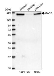
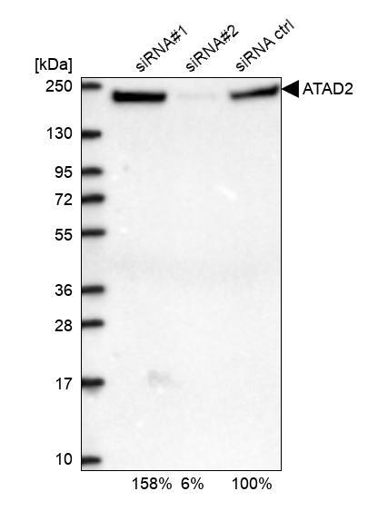
- Western blot analysis in U2OS cells transfected with control siRNA, target specific siRNA probe #1 and #2, using Anti-ATAD2 antibody. Remaining relative intensity is presented.
- Sample type
- Human
- Protocol
- Protocol
Enhanced validation
Supportive validation
- Submitted by
- 55af80e3e0991
- Enhanced method
- Genetic validation
- Main image
- Experimental details
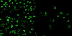
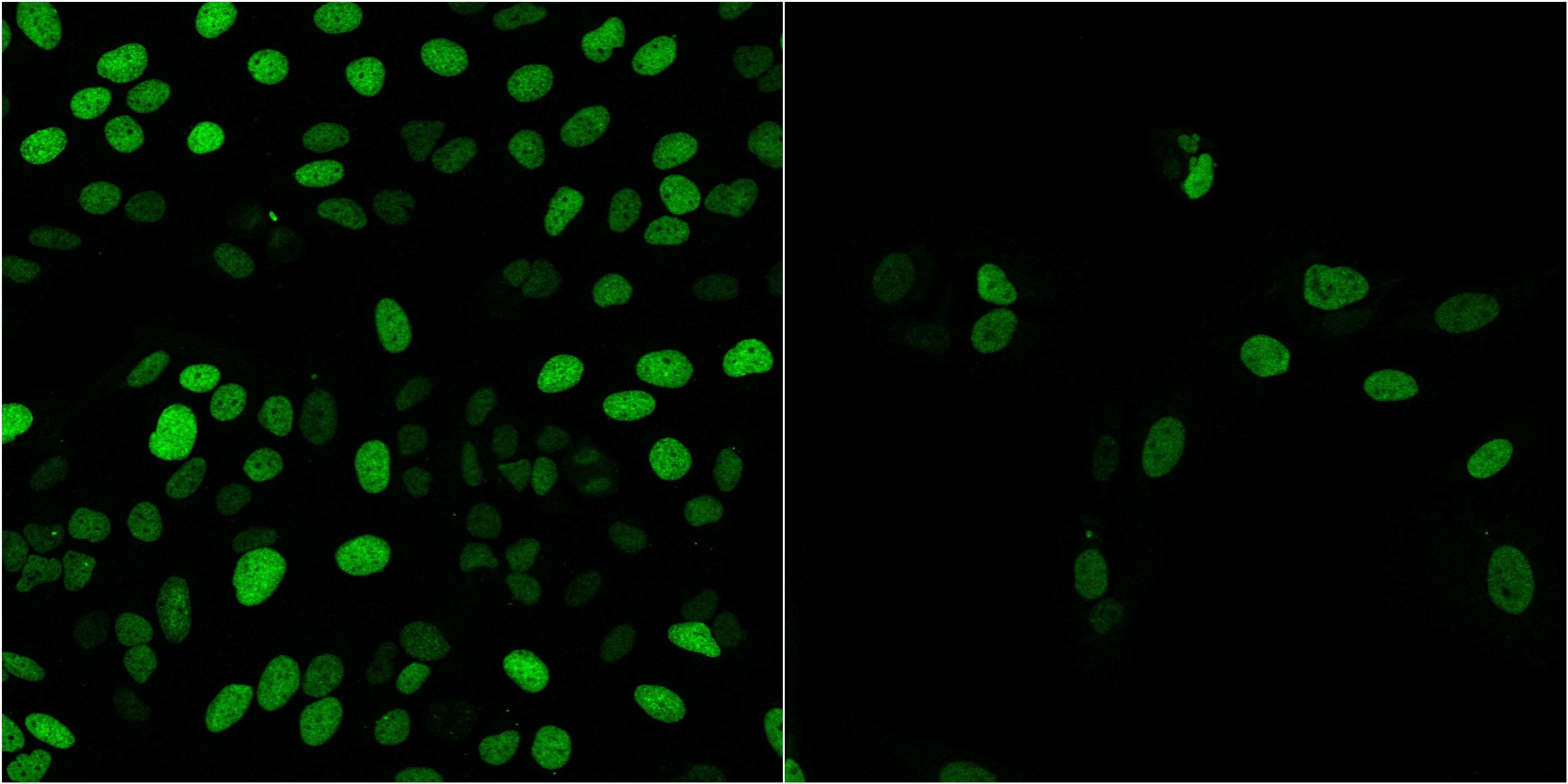
- Confocal images of immunofluorescently stained human U-2 OS cells.The protein ATAD2 is shown in green. The image to the left show cells transfected with control siRNA and the image to the right show cells where ATAD2 has been downregulated with specific siRNA.
- Sample type
- U-2 OS cells
- Primary Ab dilution
- 1:70
- Secondary Ab
- Secondary Ab
- Secondary Ab dilution
- 1:800
- Knockdown/Genetic Approaches Application
- Immunocytochemistry
Supportive validation
- Submitted by
- Atlas Antibodies (provider)
- Main image
- Experimental details
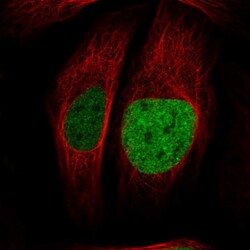
- Immunofluorescent staining of human cell line U-2 OS shows localization to nucleoplasm.
- Sample type
- Human

Supportive validation
- Submitted by
- Atlas Antibodies (provider)
- Enhanced method
- Orthogonal validation
- Main image
- Experimental details
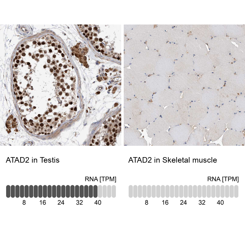
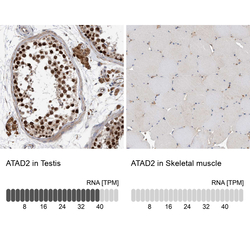
- Immunohistochemistry analysis in human testis and skeletal muscle tissues using HPA029424 antibody. Corresponding ATAD2 RNA-seq data are presented for the same tissues.
- Sample type
- Human
- Protocol
- Protocol