 Explore
 Validate
 Learn
LearnAMAb90541
antibody from Atlas Antibodies
Targeting: ATAD2
ANCCA, CT137, DKFZp667N1320, MGC29843, MGC5254, PRO2000
 Western blot
 Immunocytochemistry
Antibody data
- Antibody Data
- Antigen structure
- References [2]
- Comments [0]
- Validations
- Western blot [1]
- Immunocytochemistry [1]
- Immunohistochemistry [1]
Submit
Validation data
Reference
Comment
Report error
- Product number
- AMAb90541 - Provider product page

- Provider
- Atlas Antibodies
- Proper citation
- Atlas Antibodies Cat#AMAb90541, RRID:AB_2665580
- Product name
- Anti-ATAD2
- Antibody type
- Monoclonal
- Description
- Monoclonal Antibody against Human ATAD2, Clone ID: CL0182, Gene description: ATPase family, AAA domain containing 2, Alternative Gene Names: DKFZp667N1320, MGC29843, MGC5254, PRO2000, Validated applications: WB, IHC, ICC, Uniprot ID: Q6PL18, Storage: Store at +4°C for short term storage. Long time storage is recommended at -20°C.
- Reactivity
- Human
- Host
- Mouse
- Conjugate
- Unconjugated
- Isotype
- IgG
- Antibody clone number
- CL0182
- Vial size
- 100 µl
- Concentration
- 1.0 mg/ml
- Storage
- Store at +4°C for short term storage. Long time storage is recommended at -20°C.
- Handling
- The antibody solution should be gently mixed before use.
Submitted references ATAD2 is associated with malignant characteristics of pancreatic cancer cells
ATAD2 is highly expressed in ovarian carcinomas and indicates poor prognosis.
Liu N, Funasaka K, Obayashi T, Miyahara R, Hirooka Y, Goto H, Senga T
Oncology Letters 2019
Oncology Letters 2019
ATAD2 is highly expressed in ovarian carcinomas and indicates poor prognosis.
Wan WN, Zhang YX, Wang XM, Liu YJ, Zhang YQ, Que YH, Zhao WJ
Asian Pacific journal of cancer prevention : APJCP 2014;15(6):2777-83
Asian Pacific journal of cancer prevention : APJCP 2014;15(6):2777-83
No comments: Submit comment
Enhanced validation
- Submitted by
- Atlas Antibodies (provider)
- Enhanced method
- Genetic validation
- Main image

- Experimental details
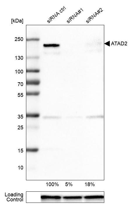
- Western blot analysis in U-251MG cells transfected with control siRNA, target specific siRNA probe #1 and #2, using Anti-ATAD2 antibody. Remaining relative intensity is presented. Loading control: Anti-GAPDH.
- Sample type
- Human
- Protocol
- Protocol
Supportive validation
- Submitted by
- Atlas Antibodies (provider)
- Main image
- Experimental details
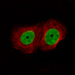
- Immunofluorescence staining of MCF7 cells using the Anti-ATAD2 monoclonal antibody, showing specific staining in nucleoplasm in green. Microtubule- and nuclear probes are visualized in red and blue, respectively (where available).
- Sample type
- Human

Supportive validation
- Submitted by
- Atlas Antibodies (provider)
- Enhanced method
- Orthogonal validation
- Main image
- Experimental details
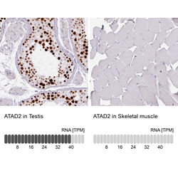
- Immunohistochemistry analysis in human testis and skeletal muscle tissues using AMAb90541 antibody. Corresponding ATAD2 RNA-seq data are presented for the same tissues.
- Sample type
- Human
- Protocol
- Protocol
