 Explore
 Validate
 Learn
Learn Western blot
 Immunocytochemistry
Antibody data
- Antibody Data
- Antigen structure
- References [0]
- Comments [0]
- Validations
- Western blot [1]
- Immunohistochemistry [4]
Submit
Validation data
Reference
Comment
Report error
- Product number
- LS-C344153 - Provider product page

- Provider
- LSBio
- Product name
- PPARA / PPAR Alpha Antibody (clone 1331CT894.186.143) LS-C344153
- Antibody type
- Monoclonal
- Description
- Protein G purified
- Reactivity
- Human, Mouse
- Host
- Mouse
- Isotype
- IgG
- Antibody clone number
- 1331CT894.186.143
- Storage
- Maintain refrigerated at 2°C to 8°C for up to 6 months. For long term storage store at -20°C.
No comments: Submit comment
Enhanced validation
- Submitted by
- LSBio (provider)
- Enhanced method
- Genetic validation
- Main image
- Experimental details
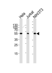
- Western blot of lysates from HeLa, Jurkat, mouse NIH/3T3 cell line (from left to right) with PPARA Antibody. Antibody was diluted at 1:1000 at each lane. A goat anti-mouse IgG H&L (HRP) at 1:3000 dilution was used as the secondary antibody. Lysates at 35 ug per lane.
Supportive validation
- Submitted by
- LSBio (provider)
- Enhanced method
- Genetic validation
- Main image
- Experimental details
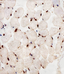
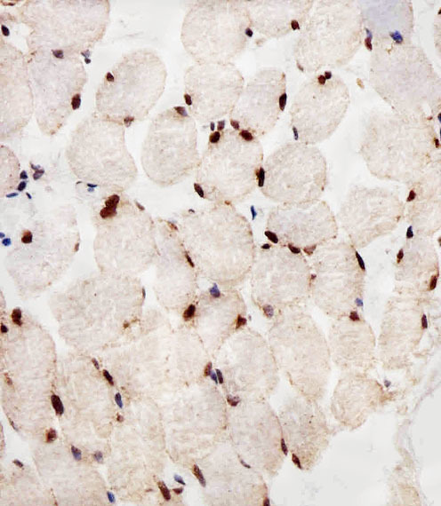
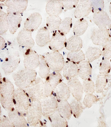
- Immunohistochemical of paraffin-embedded H. skeletal muscle section using PPARA Antibody. Antibody was diluted at 1:25 dilution. A peroxidase-conjugated goat anti-rabbit IgG at 1:400 dilution was used as the secondary antibody, followed by DAB staining.
- Submitted by
- LSBio (provider)
- Enhanced method
- Genetic validation
- Main image
- Experimental details
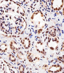
- Immunohistochemical of paraffin-embedded H. kidney section using PPARA Antibody. Antibody was diluted at 1:25 dilution. A peroxidase-conjugated goat anti-rabbit IgG at 1:400 dilution was used as the secondary antibody, followed by DAB staining.
- Submitted by
- LSBio (provider)
- Enhanced method
- Genetic validation
- Main image
- Experimental details
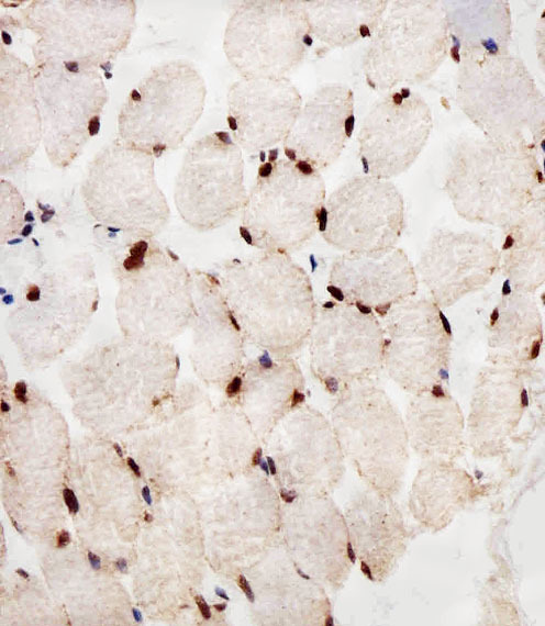
- Immunohistochemical of paraffin-embedded H. skeletal muscle section using PPARA Antibody. Antibody was diluted at 1:25 dilution. A peroxidase-conjugated goat anti-rabbit IgG at 1:400 dilution was used as the secondary antibody, followed by DAB staining.
- Submitted by
- LSBio (provider)
- Enhanced method
- Genetic validation
- Main image

- Experimental details
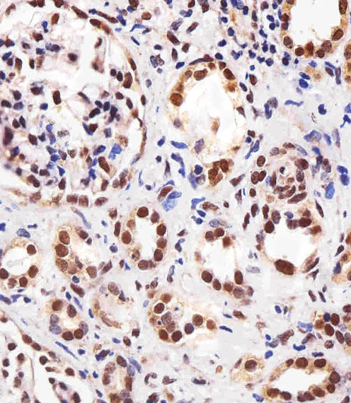
- Immunohistochemical of paraffin-embedded H. kidney section using PPARA Antibody. Antibody was diluted at 1:25 dilution. A peroxidase-conjugated goat anti-rabbit IgG at 1:400 dilution was used as the secondary antibody, followed by DAB staining.
