 Explore
 Validate
 Learn
Learn Western blot
 ELISA
ELISAAntibody data
- Antibody Data
- Antigen structure
- References [0]
- Comments [0]
- Validations
- Western blot [1]
- ELISA [3]
- Immunohistochemistry [6]
Submit
Validation data
Reference
Comment
Report error
- Product number
- LS-C745293 - Provider product page

- Provider
- LSBio
- Product name
- PPARA / PPAR Alpha Antibody (aa1-25) LS-C745293
- Antibody type
- Polyclonal
- Description
- Affinity purified
- Reactivity
- Human, Mouse
- Host
- Rabbit
- Isotype
- IgG
- Storage
- Store vial at -20°C or below prior to opening. Dilute 1:10 to minimize loss. Store the vial at -20°C or below after dilution. Avoid freeze-thaw cycles.
No comments: Submit comment
Enhanced validation
- Submitted by
- LSBio (provider)
- Enhanced method
- Genetic validation
- Main image
- Experimental details
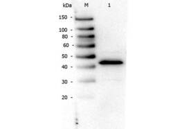
- Western Blot of rabbit anti-PPAR Alpha (N-terminal Specific) antibody. Lane 1: NIH/3T3. Load: 10 µg per lane. Primary antibody: PPAR Alpha (N-terminal specific) antibody at 1:1,000 for overnight at 4°C. Secondary antibody: Peroxidase rabbit secondary antibody at 1:40,000 for 30 min at RT. Block: Blocking Buffer for Fluorescent Western Blotting at RT for 30 min. Predicted/Observed size: ~50 kDa for PPAR Alpha.

Supportive validation
- Submitted by
- LSBio (provider)
- Enhanced method
- Genetic validation
- Main image
- Experimental details
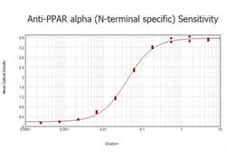
- ELISA results of purified Rabbit anti-PPAR Alpha (N-terminal specific) Antibody tested against BSA-conjugated peptide of immunizing peptide. Each well was coated in duplicate with 0.1µg of conjugate. The starting dilution of antibody was 5µg/ml and the X-axis represents the Log10 of a 3-fold dilution. This titration is a 4-parameter curve fit where the IC50 is defined as the titer of the antibody. Assay performed using 3% fish gel, Goat anti-Rabbit IgG Antibody Peroxidase Conjugated (Min X Bv Ch Gt GP Ham Hs Hu Ms Rt & Sh Serum Proteins)

- Submitted by
- LSBio (provider)
- Main image
- Experimental details
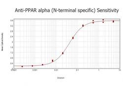
- ELISA results of purified Rabbit anti-PPAR Alpha (N-terminal specific) Antibody tested against BSA-conjugated peptide of immunizing peptide. Each well was coated in duplicate with 0.1µg of conjugate. The starting dilution of antibody was 5µg/ml and the X-axis represents the Log10 of a 3-fold dilution. This titration is a 4-parameter curve fit where the IC50 is defined as the titer of the antibody. Assay performed using 3% fish gel, Goat anti-Rabbit IgG Antibody Peroxidase Conjugated (Min X Bv Ch Gt GP Ham Hs Hu Ms Rt & Sh Serum Proteins)

- Submitted by
- LSBio (provider)
- Main image

- Experimental details
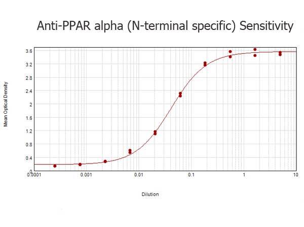
- ELISA results of purified Rabbit anti-PPAR Alpha (N-terminal specific) Antibody tested against BSA-conjugated peptide of immunizing peptide. Each well was coated in duplicate with 0.1µg of conjugate. The starting dilution of antibody was 5µg/ml and the X-axis represents the Log10 of a 3-fold dilution. This titration is a 4-parameter curve fit where the IC50 is defined as the titer of the antibody. Assay performed using 3% fish gel, Goat anti-Rabbit IgG Antibody Peroxidase Conjugated (Min X Bv Ch Gt GP Ham Hs Hu Ms Rt & Sh Serum Proteins)
Enhanced validation
- Submitted by
- LSBio (provider)
- Enhanced method
- Genetic validation
- Main image

- Experimental details
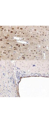
- Immunohistochemistry using the anti-PPAR antibody, showing staining of PPAR alpha in rat brain sections, highlighting cytoplasmic staining in ependymal cells and neurons in frontal cortex. Bottom image shows subventricular zone (svz) of lateral ventrical (exit point of progenitor olfactory neurones); top image shows frontal cortex in the same section. Cytoplasmic staining is also observed in the corpus callosum (bottom image) and in dendritic fields of the cortex. Formalin/PFA-fixed paraffin-embedded sections of rat brain tissue were incubated with the primary antibody at 1:200 for 1 hour. Antigen retrieval was performed by heat induction in citrate buffer pH 6.0.
- Submitted by
- LSBio (provider)
- Enhanced method
- Genetic validation
- Main image
- Experimental details
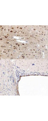
- Immunohistochemistry using the anti-PPAR antibody, showing staining of PPAR alpha in rat brain sections, highlighting cytoplasmic staining in ependymal cells and neurons in frontal cortex. Bottom image shows subventricular zone (svz) of lateral ventrical (exit point of progenitor olfactory neurones); top image shows frontal cortex in the same section. Cytoplasmic staining is also observed in the corpus callosum (bottom image) and in dendritic fields of the cortex. Formalin/PFA-fixed paraffin-embedded sections of rat brain tissue were incubated with the primary antibody at 1:200 for 1 hour. Antigen retrieval was performed by heat induction in citrate buffer pH 6.0.

- Submitted by
- LSBio (provider)
- Enhanced method
- Genetic validation
- Main image

- Experimental details
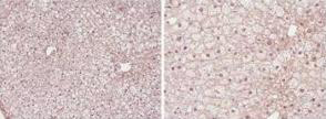
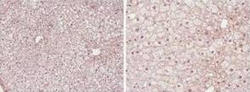
- Immunohistochemistry (Formalin/PFA-fixed paraffin-embedded sections) showing PPAR alpha antibody staining of PPAR alpha protein in mouse liver tissue section (Formalin/PFA-fixed paraffin-embedded sections). Tissue underwent formaldehyde fixation before enzymatic antigen retrieval with 0.05% protease in PBS for 5 minutes. Sample was then blocked with 5% serum for 20 minutes at 20°C. The primary antibody was diluted 1:50 and incubated with sample in Tris plus 5% normal goat serum for 1 hour at 20°C. A Biotin conjugated goat polyclonal to rabbit IgG was used at dilution at 1:500 as secondary antibody. Images show nuclear staining in hepatocytes (perfusion-fixed mouse, 10 and 40x microscope magnification).
- Submitted by
- LSBio (provider)
- Enhanced method
- Genetic validation
- Main image
- Experimental details
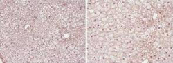
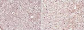
- Immunohistochemistry (Formalin/PFA-fixed paraffin-embedded sections) showing PPAR alpha antibody staining of PPAR alpha protein in mouse liver tissue section (Formalin/PFA-fixed paraffin-embedded sections). Tissue underwent formaldehyde fixation before enzymatic antigen retrieval with 0.05% protease in PBS for 5 minutes. Sample was then blocked with 5% serum for 20 minutes at 20°C. The primary antibody was diluted 1:50 and incubated with sample in Tris plus 5% normal goat serum for 1 hour at 20°C. A Biotin conjugated goat polyclonal to rabbit IgG was used at dilution at 1:500 as secondary antibody. Images show nuclear staining in hepatocytes (perfusion-fixed mouse, 10 and 40x microscope magnification).

- Submitted by
- LSBio (provider)
- Main image
- Experimental details
- Immunohistochemistry (Formalin/PFA-fixed paraffin-embedded sections) showing PPAR alpha antibody staining of PPAR alpha protein in mouse liver tissue section (Formalin/PFA-fixed paraffin-embedded sections). Tissue underwent formaldehyde fixation before enzymatic antigen retrieval with 0.05% protease in PBS for 5 minutes. Sample was then blocked with 5% serum for 20 minutes at 20°C. The primary antibody was diluted 1:50 and incubated with sample in Tris plus 5% normal goat serum for 1 hour at 20°C. A Biotin conjugated goat polyclonal to rabbit IgG was used at dilution at 1:500 as secondary antibody. Images show nuclear staining in hepatocytes (perfusion-fixed mouse, 10 and 40x microscope magnification).
- Submitted by
- LSBio (provider)
- Main image

- Experimental details
- Immunohistochemistry using the anti-PPAR antibody, showing staining of PPAR alpha in rat brain sections, highlighting cytoplasmic staining in ependymal cells and neurons in frontal cortex. Bottom image shows subventricular zone (svz) of lateral ventrical (exit point of progenitor olfactory neurones); top image shows frontal cortex in the same section. Cytoplasmic staining is also observed in the corpus callosum (bottom image) and in dendritic fields of the cortex. Formalin/PFA-fixed paraffin-embedded sections of rat brain tissue were incubated with the primary antibody at 1:200 for 1 hour. Antigen retrieval was performed by heat induction in citrate buffer pH 6.0.
