Explore
 Validate
 Learn
Learn Western blot
 Immunoprecipitation
 Immunohistochemistry
Antibody data
- Antibody Data
- Antigen structure
- References [1]
- Comments [0]
- Validations
- Western blot [6]
- Chromatin Immunoprecipitation [4]
- Other assay [3]
Submit
Validation data
Reference
Comment
Report error
- Product number
- PA5-85125 - Provider product page

- Provider
- Invitrogen Antibodies
- Product name
- PPAR alpha Polyclonal Antibody
- Antibody type
- Polyclonal
- Antigen
- Recombinant full-length protein
- Description
- Keep as concentrated solution. Predicted reactivity: Mouse (91%), Rat (88%), Dog (92%), Pig (92%), Rhesus Monkey (98%), Bovine (88%), Guinea pig (87%). Positive Control: Rat primary hepatocyte, mouse liver, mouse brain, mouse lung. Store product as a concentrated solution. Centrifuge briefly prior to opening the vial.
- Reactivity
- Human, Mouse, Rat
- Host
- Rabbit
- Isotype
- IgG
- Vial size
- 100 μL
- Concentration
- 1.44 mg/mL
- Storage
- Store at 4°C short term. For long term storage, store at -20°C, avoiding freeze/thaw cycles.
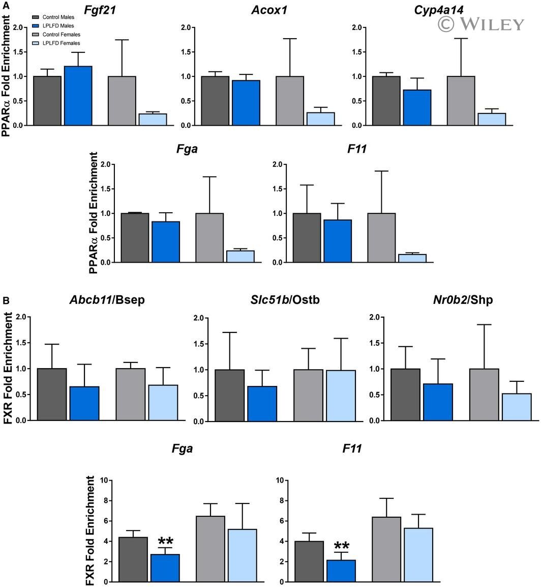
Submitted references Coagulopathy in Malnourished Mice Is Sexually Dimorphic and Regulated by Nutrient-Sensing Nuclear Receptors.
Preidis GA, Soni KG, Suh JH, Halder T, Kim KH, Choi JM, Li F, Devaraj S, Conner ME, Coarfa C, Jung SY, Moore DD
Hepatology communications 2020 Dec;4(12):1835-1850
Hepatology communications 2020 Dec;4(12):1835-1850
No comments: Submit comment
Supportive validation
- Submitted by
- Invitrogen Antibodies (provider)
- Main image
- Experimental details
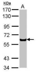
- Western blot analysis of PPAR alpha in A) rat primary hepatocyte using PPAR alpha polyclonal antibody (Product # PA5-85125) using 30 µg of sample at a dilution of 1:1000. Sample was then incubated with HRP-conjugated anti-rabbit IgG secondary antibody. Prior to incubation with primary antibody, the sample was separated on 7.5% SDS-PAGE.

- Submitted by
- Invitrogen Antibodies (provider)
- Main image
- Experimental details
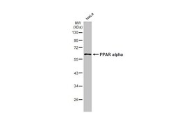
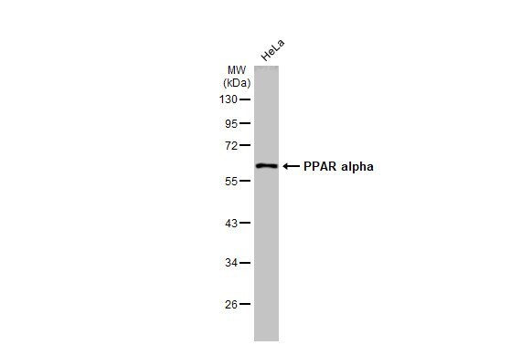
- Western Blot using PPAR alpha Polyclonal Antibody (Product # PA5-85125). Whole cell extract (30 µg) was separated by 10% SDS-PAGE, and the membrane was blotted with PPAR alpha Polyclonal Antibody (Product # PA5-85125) diluted at 1:2,000. The HRP-conjugated anti-rabbit IgG antibody was used to detect the primary antibody.
- Submitted by
- Invitrogen Antibodies (provider)
- Main image
- Experimental details
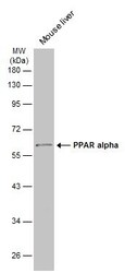
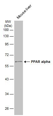
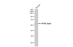
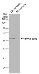
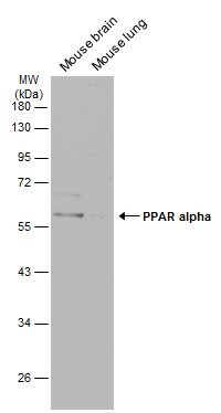
- Western blot analysis of PPAR alpha was performed by separating 50 µg of mouse tissue extract by 10% SDS-PAGE. Proteins were transferred to a membrane and probed with a PPAR alpha Polyclonal Antibody (Product # PA5-85125) at a dilution of 1:500. The HRP-conjugated anti-rabbit IgG antibody was used to detect the primary antibody.
- Submitted by
- Invitrogen Antibodies (provider)
- Main image
- Experimental details
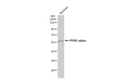
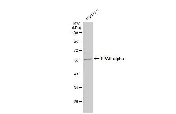
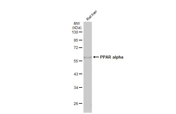
- Western blot analysis of PPAR alpha was performed by separating 50 µg of rat tissue extract by 10% SDS-PAGE. Proteins were transferred to a membrane and probed with a PPAR alpha Polyclonal Antibody (Product # PA5-85125) at a dilution of 1:500. The HRP-conjugated anti-rabbit IgG antibody was used to detect the primary antibody.
- Submitted by
- Invitrogen Antibodies (provider)
- Main image
- Experimental details
- Western blot analysis of PPAR alpha was performed by separating 50 µg of various tissue extracts by 10% SDS-PAGE. Proteins were transferred to a membrane and probed with a PPAR alpha Polyclonal Antibody (Product # PA5-85125) at a dilution of 1:500. The HRP-conjugated anti-rabbit IgG antibody was used to detect the primary antibody.
Supportive validation
- Submitted by
- Invitrogen Antibodies (provider)
- Main image

- Experimental details
- ChIP (chromatin immunoprecipitation) analysis of PPAR alpha in HepG2 chromatin extract with PPAR alpha polyclonal antibody (Product # PA5-85125) using 5 µg of sample.

- Submitted by
- Invitrogen Antibodies (provider)
- Main image

- Experimental details
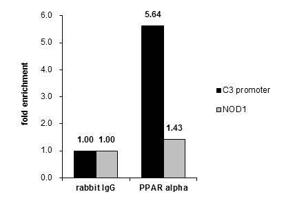
- Cross-linked ChIP was performed with HepG2 chromatin extract and 5 µg of either control rabbit IgG or PPAR alpha Polyclonal Antibody (Product # PA5-85125). The precipitated DNA was detected by PCR with primer set targeting to C3 promotor or NOD1 gene.

- Submitted by
- Invitrogen Antibodies (provider)
- Main image

- Experimental details
- ChIP (chromatin immunoprecipitation) analysis of PPAR alpha in HepG2 chromatin extract with PPAR alpha polyclonal antibody (Product # PA5-85125) using 5 µg of sample.

- Submitted by
- Invitrogen Antibodies (provider)
- Main image

- Experimental details
- Cross-linked ChIP was performed with HepG2 chromatin extract and 5 µg of either control rabbit IgG or PPAR alpha Polyclonal Antibody (Product # PA5-85125). The precipitated DNA was detected by PCR with primer set targeting to C3 promotor or NOD1 gene.
Supportive validation
- Submitted by
- Invitrogen Antibodies (provider)
- Main image

- Experimental details
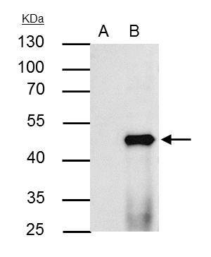
- Immunoprecipitation analysis of PPAR alpha in HepG2 whole cell lysate, A) control of pre-immune rabbit IgG, B) immunoprecipitation of PPAR alpha protein with PPAR alpha polyclonal antibody (Product # PA5-85125) using 2.5 µg of sample at a dilution of 1:1000. Sample was then incubated with HRP-conjugated anti-rabbit IgG secondary antibody. Prior to incubation with primary antibody, the sample was separated on 10% SDS-PAGE.

- Submitted by
- Invitrogen Antibodies (provider)
- Main image

- Experimental details
- PPAR alpha Polyclonal Antibody immunoprecipitates PPAR alpha protein in IP experiments. IP Sample: HepG2 whole cell lysate/extract A : Control with 2.5 µg of pre-immune rabbit IgG B : Immunoprecipitation of PPAR alpha by 2.5 µg of PPAR alpha Polyclonal Antibody (Product # PA5-85125) 10% SDS-PAGE The immunoprecipitated PPAR alpha protein was detected by PPAR alpha Polyclonal Antibody (Product # PA5-85125) diluted at 1:1,000. Anti-rabbit IgG (HRP) was used as a secondary reagent.
- Submitted by
- Invitrogen Antibodies (provider)
- Main image

- Experimental details
- 6 FIG. Altered hepatic nuclear receptor expression and FXR agonists in malnutrition. (A-C) Relative concentrations of FXR and PPARalpha protein in liver, determined by western blot, reveal a profound decrease of PPARalpha expression in malnourished male livers (n = 3-4). (D) Relative expression of PMP70 correlates directly with PPARalpha protein levels in malnourished male livers (n = 3-4) and supports qualitative evaluation by electron microscopy revealing loss of hepatocyte peroxisomes (Supporting Fig. S8). (E) Targeted bile acid mass spectrometry from liver, plasma, and stool found total fecal bile acid levels to be significantly decreased in malnourished male mice. (F) Individual stool bile acid metabolites show multiple decreased bile acids and sex specificity (n = 6). Individual liver and plasma bile acid metabolites are shown in Supporting Fig. S9. Data in (B-D) are mean + SD. Box and whisker plots (E,F) show interquartile range (box), median (vertical line), and outliers (whiskers). Student t test; **** P < 0.0001, ** P < 0.01, * P < 0.05 compared to control mice of the same sex. [Corrections added on October 24, 2020 after first online publication: the figure 6 has been replaced with the correct one.]