 Explore
 Validate
 Learn
Learn Western blot
 ELISA
ELISAAntibody data
- Antibody Data
- Antigen structure
- References [2]
- Comments [0]
- Validations
- Western blot [3]
- Immunocytochemistry [1]
- Immunohistochemistry [2]
Submit
Validation data
Reference
Comment
Report error
- Product number
- GTX28934 - Provider product page

- Provider
- GeneTex
- Proper citation
- GeneTex Cat#GTX28934, RRID:AB_372526
- Product name
- PPAR alpha antibody
- Antibody type
- Polyclonal
- Reactivity
- Human, Mouse, Rat, Bovine, Canine, Hamster, Porcine
- Host
- Rabbit
Submitted references Expression of cytochrome P450 epoxygenases and soluble epoxide hydrolase is regulated by hypolipidemic drugs in dose-dependent manner.
Effect of monomeric adiponectin on cardiac function and perfusion in anesthetized pig.
Cizkova K
Toxicology and applied pharmacology 2018 Sep 15;355:156-163
Toxicology and applied pharmacology 2018 Sep 15;355:156-163
Effect of monomeric adiponectin on cardiac function and perfusion in anesthetized pig.
Grossini E, Prodam F, Walker GE, Sigaudo L, Farruggio S, Bellofatto K, Marotta P, Molinari C, Mary D, Bona G, Vacca G
The Journal of endocrinology 2014 Jul;222(1):137-49
The Journal of endocrinology 2014 Jul;222(1):137-49
No comments: Submit comment
Supportive validation
- Submitted by
- GeneTex (provider)
- Main image
- Experimental details
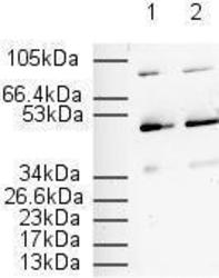
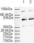
- Western Blot using GTX28934 on 20 g / lane 3T3 Whole Cell Lysate. Goat anti-rabbit IgG HRP Conjugate used as secondary at 1/2000. Exposure time: 10 mins. Lane 1: 1/500 Lane 2: 1/1000.
- Validation comment
- WB
- Submitted by
- GeneTex (provider)
- Main image

- Experimental details
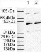
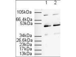
- Affinity Purified anti-PPAR alpha (N -terminal specific) (Rabbit) is shown to detect a 52 kDa band corresponding to PPAR alpha present in a 3T3 whole cell lysate. Approximately 20 g of lysate was loaded per lane for SDS-PAGE. Detection occurred after using a 1:500 (lane 1) or 1:1000 (lane 2) dilution of antibody followed by 1:2000 dilution of HRP Goat-anti-Rabbit IgG for visualization.
- Validation comment
- WB
- Submitted by
- GeneTex (provider)
- Main image
- Experimental details
- Affinity Purified anti-PPAR alpha (N -terminal specific) (Rabbit) is shown to detect a 52 kDa band corresponding to PPAR alpha present in a 3T3 whole cell lysate. Approximately 20 ?g of lysate was loaded per lane for SDS-PAGE. Detection occurred after using a 1:500 (lane 1) or 1:1000 (lane 2) dilution of antibody followed by 1:2000 dilution of HRP Goat-a-Rabbit IgG for visualization.

Supportive validation
- Submitted by
- GeneTex (provider)
- Main image
- Experimental details
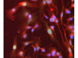
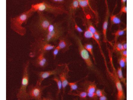
- Immunofluorescence Microscopy of Rabbit anti-PPAR alpha antibody (GTX28934). Tissue: HepG2 cells. Fixation: 4% formaldehyde fixed (10 min). Antigen retrieval: not required. Primary antibody: PPAR alpha antibody at 1 ?g/mL overnight at 4¢XC. Secondary antibody: Alexa Fluor? 488 goat anti-rabbit IgG (H+L) (green) used at a 1:1000, Alexa Fluor? 594 WGA was used to label plasma membranes (red) at a 1:200 dilution for 1h for 45 min at RT. Localization: PPAR alpha is nuclear and occasionally cytoplasmic. Staining: PPAR alpha as green fluorescent signal with DAPI (blue) nuclear counterstain.
Supportive validation
- Submitted by
- GeneTex (provider)
- Main image
- Experimental details
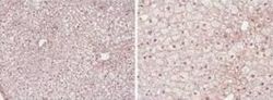
- Immunohistochemistry showing GeneTex's PPAR alpha antibody staining of PPAR alpha protein in mouse liver tissue section (Formalin/PFA-fixed paraffin-embedded sections). Tissue underwent formaldehyde fixation before enzymatic antigen retrieval with 0.05% protease in PBS for 5 minutes. Sample was then blocked with 5% serum for 20 minutes at 20¢XC. The primary antibody was diluted 1:50 and incubated with sample in Tris plus 5% normal goat serum for 1 hour at 20¢XC. A biotinylated goat polyclonal to rabbit IgG was used at dilution at 1:500 as secondary antibody. Images show nuclear staining in hepatocytes (perfusion-fixed mouse, 10 and 40x microscope magnification).
- Submitted by
- GeneTex (provider)
- Main image
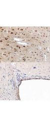
- Experimental details
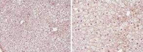
- Immunohistochemistry using GeneTex's anti-PPAR antibody, showing staining of PPAR alpha in rat brain sections, highlighting cytoplasmic staining in ependymal cells and neurons in frontal cortex. Bottom image shows subventricular zone (svz) of lateral ventrical (exit point of progenitor olfactory neurones); top image shows frontal cortex in the same section. Cytoplasmic staining is also observed in the corpus callosum (bottom image) and in dendritic fields of the cortex. Formalin/PFA-fixed paraffin-embedded sections of rat brain tissue were incubated with the primary antibody at 1:200 for 1 hour. Antigen retrieval was performed by heat induction in citrate buffer pH 6.0.
