Explore
 Validate
 Learn
Learn Western blot
 ELISA
ELISAAntibody data
- Antibody Data
- Antigen structure
- References [0]
- Comments [0]
- Validations
- Western blot [1]
- Immunohistochemistry [2]
Submit
Validation data
Reference
Comment
Report error
- Product number
- R1489P - Provider product page

- Provider
- Acris Antibodies GmbH
- Proper citation
- Acris Antibodies GmbH Cat#R1489P, RRID:AB_1006252
- Product name
- anti PPAR-alpha (N-term)
- Antibody type
- Polyclonal
- Antigen
- Synthetic peptide corresponding to amino acids 1 to 18 of Human PPAR alpha.
- Reactivity
- Human, Mouse, Rat, Bovine, Canine, Hamster
- Host
- Rabbit
- Vial size
- 0.1 mg
- Concentration
- 1.0 mg/ml (by UV absorbance at 280 nm)
No comments: Submit comment
Supportive validation
- Submitted by
- Acris Antibodies GmbH (provider)
- Main image
- Experimental details
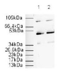
- Immunoblotting: Affinity Purified Anti-PPAR alpha (N-terminal specific) is shown to detect a 52 kDa band corresponding to Human PPAR alpha present in a 3T3 whole cell lysate. Approximately 20 µg of lysate was loaded per lane for SDS-PAGE. Detection occurred using a 1/500 (Lane 1) or 1/1000 (Lane 2) dilution of antibody followed by 1/2000 dilution of HRP Goat anti-Rabbit IgG for visualization.

Supportive validation
- Submitted by
- Acris Antibodies GmbH (provider)
- Main image

- Experimental details
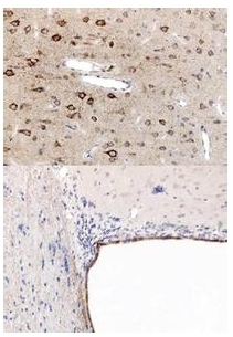
- Immunohistochemistry using PPAR antibody, showing staining of PPAR alpha in Rat brain sections, highlighting cytoplasmic staining in ependymal cells and neurons in frontal cortex. Bottom image shows subventricular zone (svz) of lateral ventrical (exit point of progenitor olfactory neurones). Top image shows frontal cortex in the same section. Cytoplasmic staining is also observed in the corpus callosum (bottom image) and in dendritic fields of the cortex. Formalin/PFA-fixed paraffin-embedded sections of rat brain tissue were incubated with the primary antibody at 1:200 for 1 hour. Antigen retrieval was performed by heat induction in citrate buffer pH 6.0.
- Submitted by
- Acris Antibodies GmbH (provider)
- Main image
- Experimental details
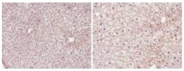
- Immunohistochemistry showing PPAR alpha antibody staining of PPAR alpha protein in Formalin/PFA-Fixed Paraffin-Embedded Mouse liver tissue section. Tissue underwent formaldehyde fixation before enzymatic antigen retrieval with 0.05% protease in PBS for 5 minutes. Sample was then blocked with 5% serum for 20 minutes at 20°C. The primary antibody was diluted 1:50 and incubated with sample in Tris plus 5% normal goat serum for 1 hour at 20°C. A Biotin conjugated Goat polyclonal to Rabbit IgG was used at dilution at 1/500 as secondary antibody. Images show nuclear staining in hepatocytes (perfusion-fixed mouse, 10 and 40x microscope magnification).