Explore
 Validate
 Learn
Learn Western blot
Antibody data
- Antibody Data
- Antigen structure
- References [0]
- Comments [0]
- Validations
- Western blot [1]
- Immunoprecipitation [1]
- Immunohistochemistry [1]
Submit
Validation data
Reference
Comment
Report error
- Product number
- MA5-55939 - Provider product page

- Provider
- Invitrogen Antibodies
- Product name
- PDE5 Monoclonal Antibody (K1E005_13D12)
- Antibody type
- Monoclonal
- Antigen
- Recombinant full-length protein
- Description
- Sequence of this protein is as follows: QSLAAAVVPS AQTLKITDFS FSDFELSDLE TALCTIRMFT DLNLVQNFQM KHEVLCRWIL SVKKNYRKNV AYHNWRHAFN TAQCMFAALK AGKIQNKLTD LEILALLIAA LSHDLDHRGV NNSYIQRSEH PLAQLYCHSI MEHHHFDQCL MILNSPGNQI LSGLSIEEYK TTLKIIKQAI LATDLALYIK RRGEFFELIR KNQFNLEDPH QKELFLAMLM TACDLSAITK PWPIQQRIAE LVATEFFDQG DRERKELNIE PTDLMNREKK NKIPSMQVGF IDAICLQLYE ALTHVSEDCF PLLDGCRKNR QKWQALAEQQ
- Reactivity
- Human
- Host
- Mouse
- Isotype
- IgG
- Antibody clone number
- K1E005_13D12
- Vial size
- 50 μg
- Concentration
- 1 mg/mL
- Storage
- Store at 4°C short term. For long term storage, store at -20°C, avoiding freeze/thaw cycles.
No comments: Submit comment
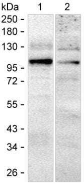
Supportive validation
- Submitted by
- Invitrogen Antibodies (provider)
- Main image
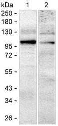
- Experimental details
- Western blot analysis of PDE5 in 15 µg of MCF7 lysate. Sample was run on 6-18% SDS-PAGE under reducing conditions, blotted onto nitrocellulose membrane, and peroxidase conjugated goat anti-mouse IgG was used as the secondary antibody. PDE5 band was visualized using ECL Substrate. Incubation with primary PDE5 monoclonal antibody (Product # MA5-55939) at a dilution of 1 µg/mL was used.
Supportive validation
- Submitted by
- Invitrogen Antibodies (provider)
- Main image
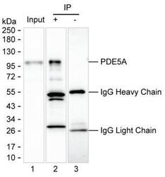
- Experimental details
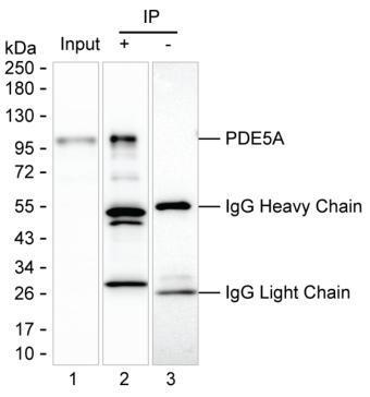
- Immunoprecipitation of PDE5 in 200 µg of HEK-293 lysate. Samples are as follows: Lane 1: HEK-293 lysate, Lane 2: PDE5 immunoprecipitated from HEK-293 lysate, Lane3: The same as Lane 2 but KT82 was used as IgG isotype control antibody. After absorption with Protein G beads, the mixture was run on 6-18% SDS-PAGE, blotted onto nitrocellulose membrane, and peroxidase conjugated goat anti-mouse IgG was used as the secondary antibody. The isotype control antibody was KT82. Incubation of samples with PDE5 monoclonal antibody (Product # MA5-55939) at a dilution of 2.5 µg was used.
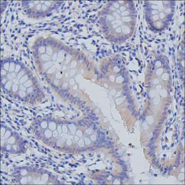
Supportive validation
- Submitted by
- Invitrogen Antibodies (provider)
- Main image
- Experimental details
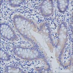
- Immunohistochemistry analysis of PDE5 in paraffin-embedded colon tissue. Sample was incubated with PDE5 monoclonal antibody (Product # MA5-55939) at a dilution of 5 µg/mL (RT, 1 hour). Antigen was retrieved through addition of boiling Tris/EDTA buffer pH 9 in a pressure cooker for 3 min. Endogenous peroxidase activity was quenched by incubating the sections with 3% H2O2 for 30 min at room temperature. Poly-peroxidase conjugated goat anti-mouse IgG was used as the secondary antibody. Diaminobenzidine was used as the chromogen. The section was counterstained with hematoxylin.