 Explore
 Validate
 Learn
Learn Western blot
Antibody data
- Antibody Data
- Antigen structure
- References [0]
- Comments [0]
- Validations
- Western blot [4]
- Immunocytochemistry [2]
- Immunohistochemistry [1]
Submit
Validation data
Reference
Comment
Report error
- Product number
- PA5-78298 - Provider product page

- Provider
- Invitrogen Antibodies
- Product name
- SETD1A Polyclonal Antibody
- Antibody type
- Polyclonal
- Antigen
- Recombinant full-length protein
- Description
- Predicted Reactivity: Bovine (90%) Store product as a concentrated solution. Centrifuge briefly prior to opening the vial.
- Reactivity
- Human, Mouse
- Host
- Rabbit
- Isotype
- IgG
- Vial size
- 100 µL
- Concentration
- 1.18 mg/mL
- Storage
- Store at 4°C short term. For long term storage, store at -20°C, avoiding freeze/thaw cycles.
No comments: Submit comment
Supportive validation
- Submitted by
- Invitrogen Antibodies (provider)
- Main image
- Experimental details
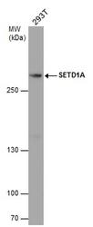
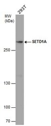
- Western blot analysis of SETD1A in whole cell lysate using 30 µg of protein. Samples were separated with 5% SDS-PAGE and incubated with SETD1A polyclonal antibody (Product # PA5-78298) using a dilution of 1:1000.
- Submitted by
- Invitrogen Antibodies (provider)
- Main image
- Experimental details
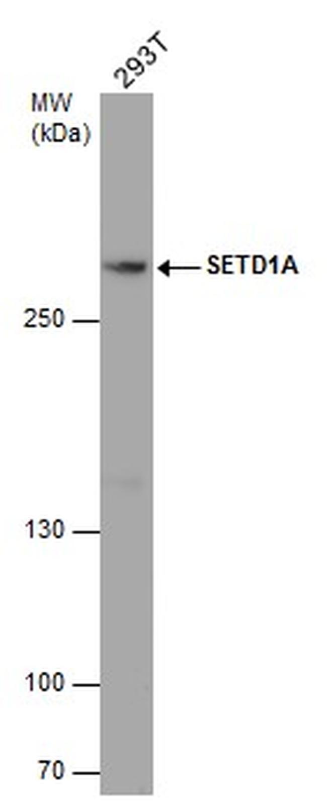
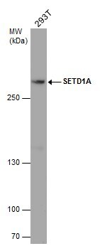
- Western Blot using SETD1A Polyclonal Antibody (Product # PA5-78298). Whole cell extract (30 µg) was separated by 5% SDS-PAGE, and the membrane was blotted with SETD1A Polyclonal Antibody (Product # PA5-78298) diluted at 1:1,000.
- Submitted by
- Invitrogen Antibodies (provider)
- Main image
- Experimental details
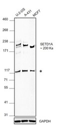
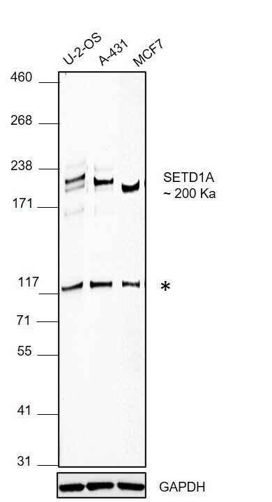
- Western blot was performed using Anti-SETD1A Polyclonal Antibody (Product # PA5-78298) and a 200 kDa band corresponding to SETD1A was observed across all the cell lines tested along with an uncharacterized band around 117 kDa. Whole cell extracts (30 µg lysate) of U-2-OS (Lane 1), A-431 (Lane 2) and MCF7 (Lane 3) were electrophoresed using NuPAGE™ 3-8% Tris-Acetate Protein Gel (Product # EA0378BOX). Resolved proteins were then transferred onto a nitrocellulose membrane (Product # IB23002) by iBlot® 2 Dry Blotting System (Product # IB21001). The blot was probed with the primary antibody (1:1000 dilution) and detected by chemiluminescence with Goat anti-Rabbit IgG (H+L) Superclonal™ Recombinant Secondary Antibody, HRP (Product # A27036,1:10000 dilution) using the iBright™ FL1500 Imaging System (Product # A44115). Chemiluminescent detection was performed using SuperSignal™ West Pico PLUS Chemiluminescent Substrate (Product # 34580).
- Submitted by
- Invitrogen Antibodies (provider)
- Main image
- Experimental details
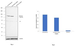
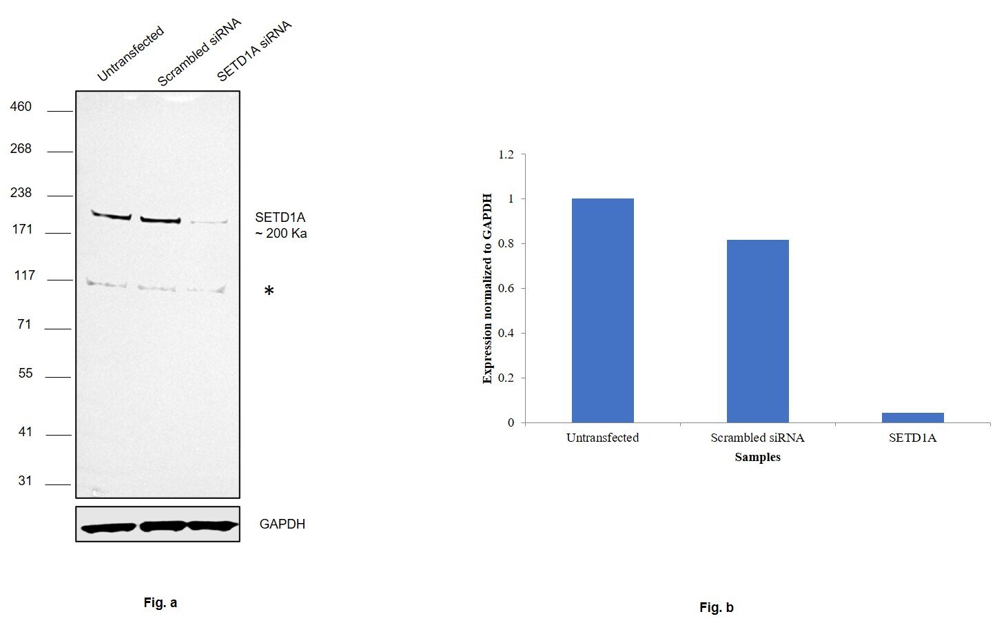
- Knockdown of SETD1A was achieved by transfecting A-431 cells with SETD1A specific siRNAs (Silencer® select Product # S18789, S18788). Western blot analysis (Fig. a) was performed using Whole cell extracts from the SETD1A knockdown cells (lane 3), non-targeting scrambled siRNA transfected cells (lane 2) and untransfected cells (lane 1), (*) representing uncharacterized bands observed. The blot was probed with SETD1A Polyclonal Antibody (Product # PA5-78298, 1:1000 dilution) and Goat anti-Rabbit IgG (H+L) Superclonal™ Recombinant Secondary Antibody, HRP (Product # A27036, 1:4000 dilution). Densitometric analysis of this western blot is shown in histogram (Fig. b). Decrease in signal upon siRNA mediated knock down confirms that antibody is specific to SETD1A.
Supportive validation
- Submitted by
- Invitrogen Antibodies (provider)
- Main image
- Experimental details
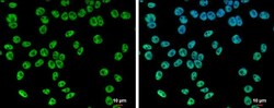
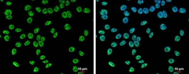
- SETD1A Polyclonal Antibody detects SETD1A protein at nucleus by immunofluorescent analysis. Sample: 293T cells were fixed in 4% paraformaldehyde at RT for 15 min. Green: SETD1A protein stained by SETD1A Polyclonal Antibody (Product # PA5-78298) diluted at 1:500. Blue: Hoechst 33342 staining. Scale bar = 10 µm.
- Submitted by
- Invitrogen Antibodies (provider)
- Main image
- Experimental details
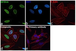
- Immunofluorescence analysis of SETD1A was performed using 70% confluent log phase HeLa cells. The cells were fixed with 4% paraformaldehyde for 15 minutes, permeabilized with 0.1% Triton™ X-100 for 10 minutes, and blocked with 2% BSA for 45 minutes at room temperature. The cells were labeled with SETD1A Polyclonal Antibody (Product # PA5-78298) at 1:100 dilution in 0.1% BSA, incubated at 4 degree celsius overnight and then labeled with Goat anti-Rabbit IgG (H+L) Superclonal™ Recombinant Secondary Antibody, Alexa Fluor® 488 conjugate (Product # A27034), (1:2000 dilution), for 45 minutes at room temperature (Panel a: Green). Nuclei (Panel b:Blue) were stained with ProLong™ Diamond Antifade Mountant with DAPI (Product # P36962). F-actin (Panel c: Red) was stained with Rhodamine Phalloidin (Product # R415,1:300 dilution). Panel d represents the merged image showing nuclear localization. Panel e represents control cells with no primary antibody to assess background. The images were captured at 60X magnification.

Supportive validation
- Submitted by
- Invitrogen Antibodies (provider)
- Main image
- Experimental details
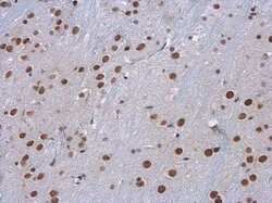
- SETD1A Polyclonal Antibody detects SETD1A protein at nucleus in mouse brain by immunohistochemical analysis. Sample: Paraffin-embedded mouse brain. SETD1A Polyclonal Antibody (Product # PA5-78298) diluted at 1:500. Antigen Retrieval: Citrate buffer, pH 6.0, 15 min.
