Explore
 Validate
 Learn
Learn Flow cytometry
Antibody data
- Antibody Data
- Antigen structure
- References [10]
- Comments [0]
- Validations
- Flow cytometry [1]
- Other assay [3]
Submit
Validation data
Reference
Comment
Report error
- Product number
- 12-1178-42 - Provider product page

- Provider
- Invitrogen Antibodies
- Product name
- CD117 (c-Kit) Monoclonal Antibody (104D2), PE, eBioscience™
- Antibody type
- Monoclonal
- Antigen
- Other
- Description
- Description: The 104D2 monoclonal antibody reacts with human CD117, also known as c-Kit , Steel factor receptor and stem cell factor receptor. A member of the tyrosine kinase receptor family, this 145 kDa molecule is expressed by hematopoietic progenitor cell subsets and mast cells. The interaction of c-Kit and Steel factor promotes proliferation and differentiation of hematopoietic progenitor cells and mast cell differentiation. CD117 is also expressed by melanocytes and plays a role in signaling and activation of these cells. Applications Reported: This 104D2 antibody has been reported for use in flow cytometric analysis. Applications Tested: This 104D2 antibody has been pre-titrated and tested by flow cytometric analysis of normal human peripheral blood cells. This can be used at 5 µL (0.25 µg) per test. A test is defined as the amount (µg) of antibody that will stain a cell sample in a final volume of 100 µL. Cell number should be determined empirically but can range from 10^5 to 10^8 cells/test. Excitation: 488-561 nm; Emission: 578 nm; Laser: Blue Laser, Green Laser, Yellow-Green Laser. Filtration: 0.2 µm post-manufacturing filtered.
- Reactivity
- Human
- Host
- Mouse
- Conjugate
- Yellow dye
- Isotype
- IgG
- Antibody clone number
- 104D2
- Vial size
- 100 Tests
- Concentration
- 5 µL/Test
- Storage
- 4° C, store in dark, DO NOT FREEZE!
Submitted references Airway and Lung Organoids from Human-Induced Pluripotent Stem Cells Can Be Used to Assess CFTR Conductance.
STS1 and STS2 Phosphatase Inhibitor Baicalein Enhances the Expansion of Hematopoietic and Progenitor Stem Cells and Alleviates 5-Fluorouracil-Induced Myelosuppression.
IL6Myc mouse is an immunocompetent model for the development of aggressive multiple myeloma.
Pharmacological inhibition of LSD1 triggers myeloid differentiation by targeting GSE1 oncogenic functions in AML.
Production and cryopreservation of definitive endoderm from human pluripotent stem cells under defined and scalable culture conditions.
Chemically-Defined, Xeno-Free, Scalable Production of hPSC-Derived Definitive Endoderm Aggregates with Multi-Lineage Differentiation Potential.
Mechanisms of Progression of Myeloid Preleukemia to Transformed Myeloid Leukemia in Children with Down Syndrome.
IL1RAP potentiates multiple oncogenic signaling pathways in AML.
Induced pluripotent stem cell-based mapping of β-globin expression throughout human erythropoietic development.
PPAR-α and glucocorticoid receptor synergize to promote erythroid progenitor self-renewal.
Demchenko A, Kondrateva E, Tabakov V, Efremova A, Salikhova D, Bukharova T, Goldshtein D, Balyasin M, Bulatenko N, Amelina E, Lavrov A, Smirnikhina S
International journal of molecular sciences 2023 Mar 27;24(7)
International journal of molecular sciences 2023 Mar 27;24(7)
STS1 and STS2 Phosphatase Inhibitor Baicalein Enhances the Expansion of Hematopoietic and Progenitor Stem Cells and Alleviates 5-Fluorouracil-Induced Myelosuppression.
Li N, Wang Y, Wang A, Zhang J, Jia C, Yu C, Song Z, Wang S, Liu L, Yi J, Bao Y, Huang Y, Sun L
International journal of molecular sciences 2023 Feb 3;24(3)
International journal of molecular sciences 2023 Feb 3;24(3)
IL6Myc mouse is an immunocompetent model for the development of aggressive multiple myeloma.
Pisano MD, Sun F, Cheng Y, Parashar D, Zhou V, Jing X, Sompallae R, Abrudan J, Zimmermann MT, Mathison A, Janz S, Pufall MA
Haematologica 2023 Dec 1;108(12):3372-3383
Haematologica 2023 Dec 1;108(12):3372-3383
Pharmacological inhibition of LSD1 triggers myeloid differentiation by targeting GSE1 oncogenic functions in AML.
Nicosia L, Boffo FL, Ceccacci E, Conforti F, Pallavicini I, Bedin F, Ravasio R, Massignani E, Somervaille TCP, Minucci S, Bonaldi T
Oncogene 2022 Feb;41(6):878-894
Oncogene 2022 Feb;41(6):878-894
Production and cryopreservation of definitive endoderm from human pluripotent stem cells under defined and scalable culture conditions.
Sahabian A, Dahlmann J, Martin U, Olmer R
Nature protocols 2021 Mar;16(3):1581-1599
Nature protocols 2021 Mar;16(3):1581-1599
Chemically-Defined, Xeno-Free, Scalable Production of hPSC-Derived Definitive Endoderm Aggregates with Multi-Lineage Differentiation Potential.
Sahabian A, Sgodda M, Naujok O, Dettmer R, Dahlmann J, Manstein F, Cantz T, Zweigerdt R, Martin U, Olmer R
Cells 2019 Dec 4;8(12)
Cells 2019 Dec 4;8(12)
Mechanisms of Progression of Myeloid Preleukemia to Transformed Myeloid Leukemia in Children with Down Syndrome.
Labuhn M, Perkins K, Matzk S, Varghese L, Garnett C, Papaemmanuil E, Metzner M, Kennedy A, Amstislavskiy V, Risch T, Bhayadia R, Samulowski D, Hernandez DC, Stoilova B, Iotchkova V, Oppermann U, Scheer C, Yoshida K, Schwarzer A, Taub JW, Crispino JD, Weiss MJ, Hayashi Y, Taga T, Ito E, Ogawa S, Reinhardt D, Yaspo ML, Campbell PJ, Roberts I, Constantinescu SN, Vyas P, Heckl D, Klusmann JH
Cancer cell 2019 Aug 12;36(2):123-138.e10
Cancer cell 2019 Aug 12;36(2):123-138.e10
IL1RAP potentiates multiple oncogenic signaling pathways in AML.
Mitchell K, Barreyro L, Todorova TI, Taylor SJ, Antony-Debré I, Narayanagari SR, Carvajal LA, Leite J, Piperdi Z, Pendurti G, Mantzaris I, Paietta E, Verma A, Gritsman K, Steidl U
The Journal of experimental medicine 2018 Jun 4;215(6):1709-1727
The Journal of experimental medicine 2018 Jun 4;215(6):1709-1727
Induced pluripotent stem cell-based mapping of β-globin expression throughout human erythropoietic development.
Vanuytsel K, Matte T, Leung A, Naing ZH, Morrison T, Chui DHK, Steinberg MH, Murphy GJ
Blood advances 2018 Aug 14;2(15):1998-2011
Blood advances 2018 Aug 14;2(15):1998-2011
PPAR-α and glucocorticoid receptor synergize to promote erythroid progenitor self-renewal.
Lee HY, Gao X, Barrasa MI, Li H, Elmes RR, Peters LL, Lodish HF
Nature 2015 Jun 25;522(7557):474-7
Nature 2015 Jun 25;522(7557):474-7
No comments: Submit comment
Supportive validation
- Submitted by
- Invitrogen Antibodies (provider)
- Main image
- Experimental details
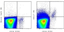
- Staining of normal human peripheral blood cells with Anti-Human CD34 FITC (Product # 11-0349-42) and Mouse IgG1 K Isotype Control PE (Product # 12-4714-81) (left) or Anti-Human CD117 (c-Kit) PE (right). CD45 dim cells in the lymphocyte gate were used for analysis.
- Conjugate
- Yellow dye

Supportive validation
- Submitted by
- Invitrogen Antibodies (provider)
- Main image
- Experimental details
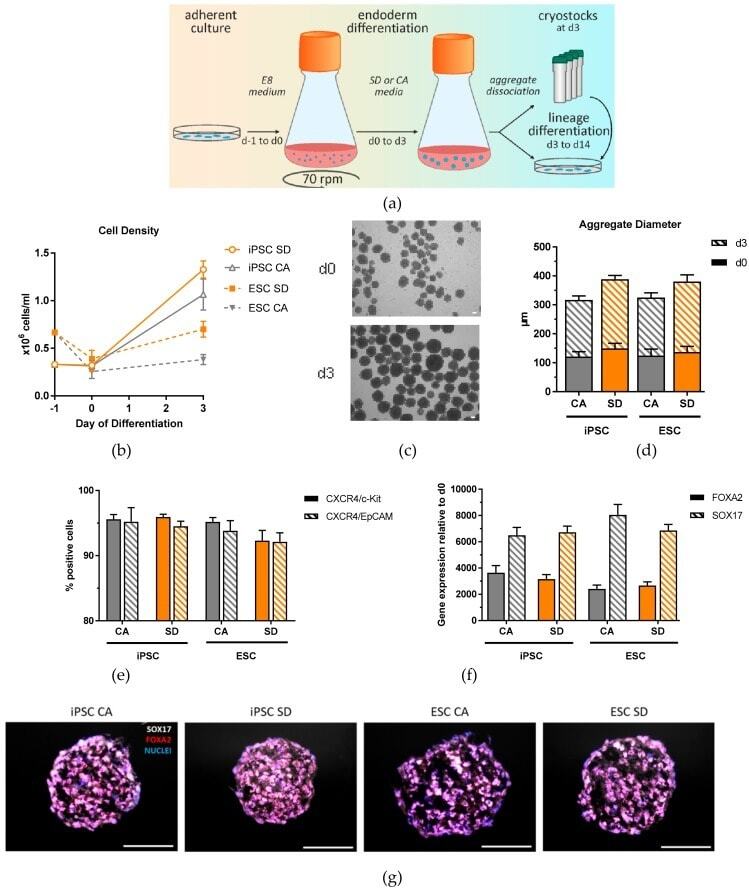
- Figure 1 Generation and characterization of DE aggregates in Erlenmeyer flasks. ( a ) Schematic overview of definitive endoderm differentiation in suspension. ( b ) Cell density analyzed at day 0 and day 3 of differentiation (n = 4-6). ( c ) Representative brightfield image of iPSC aggregates at day 0 and day 3 of differentiation (n = 4-6). ( d ) Aggregate diameters measured at day 0 and day 3 of differentiation. ( e ) Definitive endoderm efficiency quantification based on flow cytometry analysis of CXCR4/c-Kit and CXCR4/EpCAM double positive cells at day 3 of differentiation (n = 12-15). ( f ) qRT-PCR analysis of FOXA2 and SOX17 expression at day 3 of differentiation (n = 6-9). ( g ) Immunostaining of SOX17 (white) and FOXA2 (red), and nuclear stain DAPI (blue) of DE aggregates at day 3 of differentiation (n = 3). Scale bar, 100 mum. Each value of gene expression was first normalized to the reference genes and then to day 0 undifferentiated cells. Values are represented as the mean +- SEM. All n values correspond to independent experiments.
- Conjugate
- Yellow dye
- Submitted by
- Invitrogen Antibodies (provider)
- Main image
- Experimental details
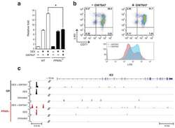
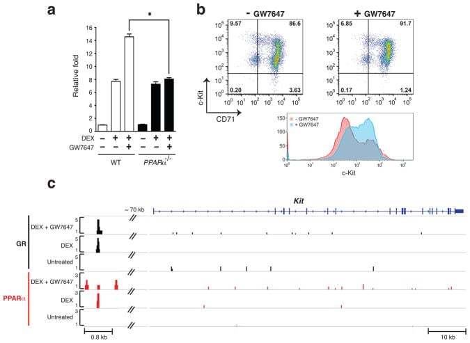
- Extended Figure 7 a, Real-time RT-PCR analysis of Kit gene expression in wild-type and PPARalpha -/- mouse BFU-E cells untreated or treated with DEX with or without addition of GW7647 (* p < 0.05; Student t test. Error bars represent mean +- S.D. from three independent experiments.); b , Human CD34+ cells were treated with or without GW7647 as described in the legend to Figure 2 . (top) At day 9 of culture, cell surface KIT and CD71 expression were analyzed by flow cytometry. (bottom) A representative histogram of KIT expression in cells treated or untreated with GW7647; c , ChIP-Seq occupancy signal map of GR and PPARalpha across the Kit locus in BFU-E cells.
- Conjugate
- Yellow dye
- Submitted by
- Invitrogen Antibodies (provider)
- Main image

- Experimental details
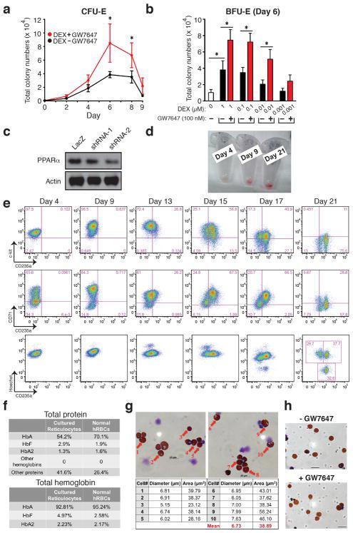
- Extended Figure 2 Human CD34 + Erythroid Differentiation System a , Total CFU-E colonies formed during day 0-9. CFU-E colony numbers were quantified by plating 1000 cells from various time points during day 0-9 of the human CD34 + erythroid culture on methylcellulose. CFU-E colonies were quantified after 12-14 days. Total CFU-E colony numbers in culture under conditions without GW7647 (black line) or with GW7647 (red line) were calculated using the total cell numbers at corresponding time points in Figure 2a. b , Human CD34+ cells were treated at day 1 with 100 nM GW7647 with or without DEX at the concentration indicated in the figure. At day 6, total cell numbers were counted and cells were collected for BFU-E colony assays. c , Protein expression of PPARalpha demonstrating shRNA knock-down efficiency via lentiviral transduction. LacZ shRNA is used as a control. shRNA-1 and -2 are both specific for PPARalpha . shRNA-2 has higher knock-down efficiency. d , Cell pellets of 1 million cells demonstrating hemoglobin accumulation during the differentiation process. e , Flow cytometry analyses of erythroid markers during the 21-day human CD34 + erythroid culture. (top row) c-kit vs. CD235a; (middle row) CD71 vs. CD235a. Note the sequential induction of c-kit, CD71 and CD235a, as well as the sequential down-regulation of c-kit and CD71, (bottom row) Enucleated reticulocytes are CD235a + Hoechst - , nuclei are CD235a - Hoechst + , and nucleated erythroblasts are CD235a + Hoechst + .
- Conjugate
- Yellow dye