Explore
 Validate
 Learn
Learn Other assay
Antibody data
- Antibody Data
- Antigen structure
- References [11]
- Comments [0]
- Validations
- Other assay [7]
Submit
Validation data
Reference
Comment
Report error
- Product number
- 14-1179-82 - Provider product page

- Provider
- Invitrogen Antibodies
- Product name
- CD117 (c-Kit) Monoclonal Antibody (YB5.B8), eBioscience™
- Antibody type
- Monoclonal
- Antigen
- Other
- Description
- Description: The YB5.B8 monoclonal antibody reacts with human CD117, also known as c-Kit , Steel factor receptor and stem cell factor receptor. A member of the tyrosine kinase receptor family, this 145 kDa molecule is expressed by hematopoietic progenitor cell subsets and mast cells. The interaction of c-Kit and Steel factor promotes proliferation and differentiation of hematopoietic progenitor cells and mast cell differentiation. CD117 is also expressed by melanocytes and plays a role in signaling and activation of these cells.
- Antibody clone number
- YB5.B8
- Concentration
- 0.5 mg/mL
Submitted references JAK2/IDH-mutant-driven myeloproliferative neoplasm is sensitive to combined targeted inhibition.
Mast cells derived from human induced pluripotent stem cells are useful for allergen tests.
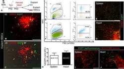
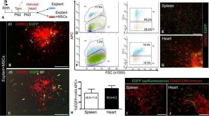
Stimulatory Effects of Mesenchymal Stem Cells on cKit+ Cardiac Stem Cells Are Mediated by SDF1/CXCR4 and SCF/cKit Signaling Pathways.
Model of fibrolamellar hepatocellular carcinomas reveals striking enrichment in cancer stem cells.
Immunophenotypic comparison of heterogenous non-sorted versus sorted mononuclear cells from human umbilical cord blood: a novel cell enrichment approach.
Human primordial germ cell commitment in vitro associates with a unique PRDM14 expression profile.
Transcriptional profile of tuberculosis antigen-specific T cells reveals novel multifunctional features.
Neoangiogenesis contributes to the development of hemophilic synovitis.
Epitope mapping and functional studies with three monoclonal antibodies to the c-kit receptor tyrosine kinase, YB5.B8, 17F11, and SR-1.
Assessment of the anti-c-kit monoclonal antibody YB5.B8 in affinity magnetic enrichment of human lung mast cells.
Monoclonal antibody YB5.B8 identifies the human c-kit protein product.
McKenney AS, Lau AN, Somasundara AVH, Spitzer B, Intlekofer AM, Ahn J, Shank K, Rapaport FT, Patel MA, Papalexi E, Shih AH, Chiu A, Freinkman E, Akbay EA, Steadman M, Nagaraja R, Yen K, Teruya-Feldstein J, Wong KK, Rampal R, Vander Heiden MG, Thompson CB, Levine RL
The Journal of clinical investigation 2018 Feb 1;128(2):789-804
The Journal of clinical investigation 2018 Feb 1;128(2):789-804
Mast cells derived from human induced pluripotent stem cells are useful for allergen tests.
Igarashi A, Ebihara Y, Kumagai T, Hirai H, Nagata K, Tsuji K
Allergology international : official journal of the Japanese Society of Allergology 2018 Apr;67(2):234-242
Allergology international : official journal of the Japanese Society of Allergology 2018 Apr;67(2):234-242
Stimulatory Effects of Mesenchymal Stem Cells on cKit+ Cardiac Stem Cells Are Mediated by SDF1/CXCR4 and SCF/cKit Signaling Pathways.
Hatzistergos KE, Saur D, Seidler B, Balkan W, Breton M, Valasaki K, Takeuchi LM, Landin AM, Khan A, Hare JM
Circulation research 2016 Sep 30;119(8):921-30
Circulation research 2016 Sep 30;119(8):921-30
Model of fibrolamellar hepatocellular carcinomas reveals striking enrichment in cancer stem cells.
Oikawa T, Wauthier E, Dinh TA, Selitsky SR, Reyna-Neyra A, Carpino G, Levine R, Cardinale V, Klimstra D, Gaudio E, Alvaro D, Carrasco N, Sethupathy P, Reid LM
Nature communications 2015 Oct 6;6:8070
Nature communications 2015 Oct 6;6:8070
Immunophenotypic comparison of heterogenous non-sorted versus sorted mononuclear cells from human umbilical cord blood: a novel cell enrichment approach.
Indumathi S, Harikrishnan R, Rajkumar JS, Dhanasekaran M
Cytotechnology 2015 Jan;67(1):107-14
Cytotechnology 2015 Jan;67(1):107-14
Human primordial germ cell commitment in vitro associates with a unique PRDM14 expression profile.
Sugawa F, Araúzo-Bravo MJ, Yoon J, Kim KP, Aramaki S, Wu G, Stehling M, Psathaki OE, Hübner K, Schöler HR
The EMBO journal 2015 Apr 15;34(8):1009-24
The EMBO journal 2015 Apr 15;34(8):1009-24
Transcriptional profile of tuberculosis antigen-specific T cells reveals novel multifunctional features.
Arlehamn CL, Seumois G, Gerasimova A, Huang C, Fu Z, Yue X, Sette A, Vijayanand P, Peters B
Journal of immunology (Baltimore, Md. : 1950) 2014 Sep 15;193(6):2931-40
Journal of immunology (Baltimore, Md. : 1950) 2014 Sep 15;193(6):2931-40
Neoangiogenesis contributes to the development of hemophilic synovitis.
Acharya SS, Kaplan RN, Macdonald D, Fabiyi OT, DiMichele D, Lyden D
Blood 2011 Feb 24;117(8):2484-93
Blood 2011 Feb 24;117(8):2484-93
Epitope mapping and functional studies with three monoclonal antibodies to the c-kit receptor tyrosine kinase, YB5.B8, 17F11, and SR-1.
Ashman LK, Bühring HJ, Aylett GW, Broudy VC, Müller C
Journal of cellular physiology 1994 Mar;158(3):545-54
Journal of cellular physiology 1994 Mar;158(3):545-54
Assessment of the anti-c-kit monoclonal antibody YB5.B8 in affinity magnetic enrichment of human lung mast cells.
Okayama Y, Hunt TC, Kassel O, Ashman LK, Church MK
Journal of immunological methods 1994 Mar 10;169(2):153-61
Journal of immunological methods 1994 Mar 10;169(2):153-61
Monoclonal antibody YB5.B8 identifies the human c-kit protein product.
Lerner NB, Nocka KH, Cole SR, Qiu FH, Strife A, Ashman LK, Besmer P
Blood 1991 May 1;77(9):1876-83
Blood 1991 May 1;77(9):1876-83
No comments: Submit comment
Supportive validation
- Submitted by
- Invitrogen Antibodies (provider)
- Main image
- Experimental details
- NULL

- Submitted by
- Invitrogen Antibodies (provider)
- Main image
- Experimental details
- NULL
- Submitted by
- Invitrogen Antibodies (provider)
- Main image
- Experimental details
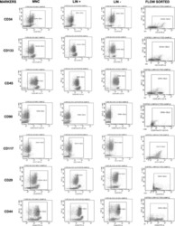
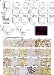
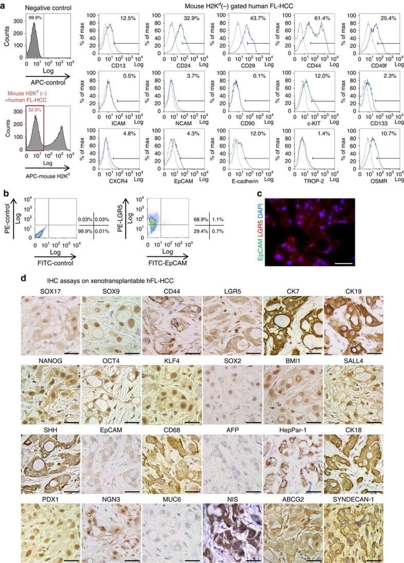
- Figure 2 Characterization of a transplantable hFL-HCC tumour line. ( a ) Representative flow cytometric findings of hFL-HCC cells, gated-mouse-H2K d -negative, were done. Antigens expressed by a significant percentage of the cells included CD44 (61.4%); CD49f (25.4%); CD24 (32.9%); CD13 (12.5%); c-KIT (12.0%); E-cadherin (12.0%); and OSMR (10.7%). Other antigens found routinely but in a smaller percentage of cells included CXCR4 (4.8%); EpCAM (4.3%); CD133 (2.3%); TROP-2 (1.4%); and ICAM1 (0.5%). ( b ) Double staining of LGR5 and EpCAM in hFL-HCC cells. LGR5+ cells accounted for 68.9% of the cells in the tumours. Of these, only 1.1% were also EpCAM+. ( c ) Immunofluorescence assay on hFL-HCC xenotransplantable tumour demonstrated strong expression of LGR5 and an absence of EpCAM. ( d ) IHC assays on the xenotransplantable tumour line. The survey included assays for endodermal stem/progenitor transcription factors and markers (SOX17, SOX9, CD44, LGR5 and CK19); pluripotency genes (NANOG, OCT4, KLF4, SOX2 and BMI1); hepatic and other markers (HepPar-1, CK18, CK7, SHH and CD68); and pancreatic/endocrine markers (PDX1, NGN3 and NIS), which were strongly expressed. EpCAM was essentially negative, and alpha-fetoprotein was negative. The scale bar, 25 mum ( c , d ).
- Submitted by
- Invitrogen Antibodies (provider)
- Main image
- Experimental details
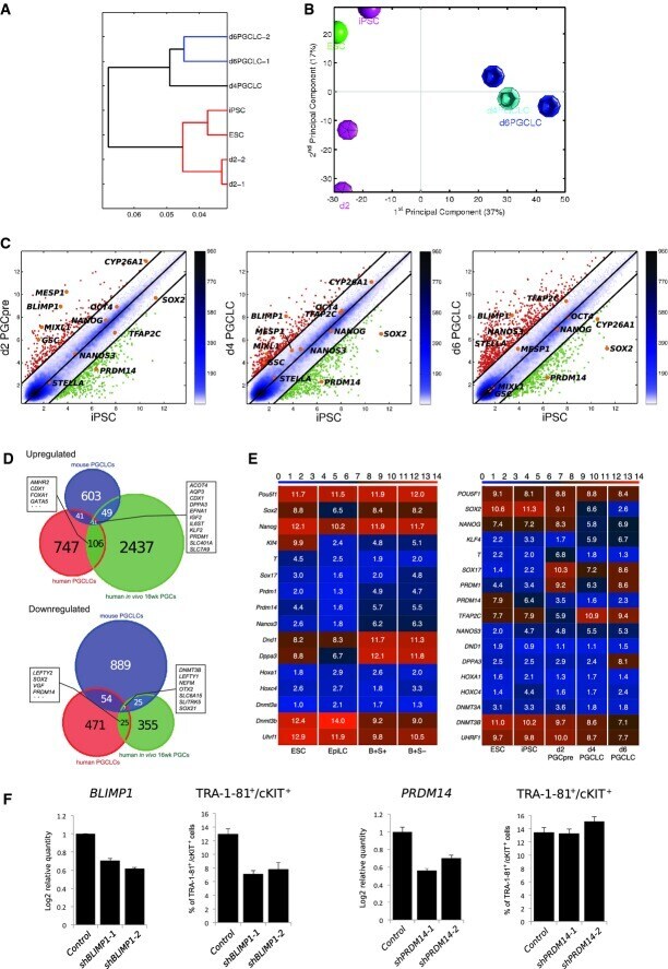
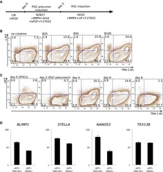
- Figure 2 Induction of PGCLCs from human iPSCs Schematic presentation of PGC-precursor and PGCLC induction. FACS analysis of the concentration-dependent effect of BMP4 on TRA-1-81 + /cKIT + PGCLCs on day 4. B25: 25 ng/ml BMP4; B50: 50 ng/ml BMP4; B100: 100 ng/ml BMP4. FACS analysis of TRA-1-81 and cKIT during PGC induction of up to day 8. Gene expression analysis of selected PGC markers in TRA-1-81 + /cKIT + and SSEA1 + /cKIT + FACS fractions of d4 PGCLCs. Samples were calibrated with iPSC values, and iPSC values depict 1. Data information: Data are presented as means +- SD ( n = 3).

- Submitted by
- Invitrogen Antibodies (provider)
- Main image
- Experimental details
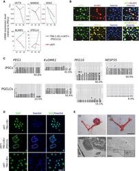
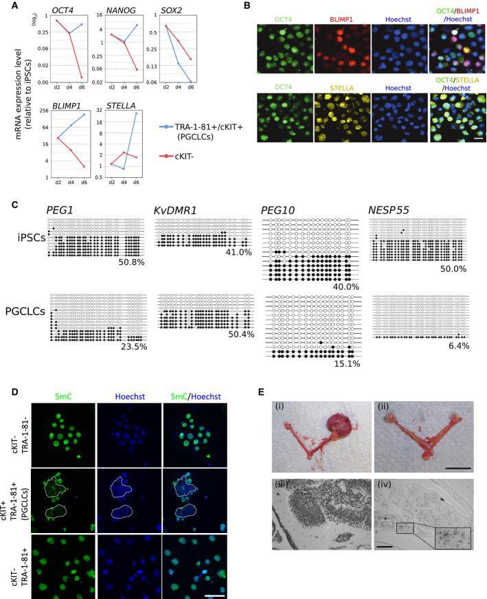
- Figure 3 Characterization of PGCLCs Gene expression dynamics of selected pluripotency and PGC genes in FACS-sorted, specified cells during PGC induction. The value for iPSCs is set as 1, and values are on log 2 scale. Immunofluorescence analysis for OCT4 (green), BLIMP1 (red), and STELLA (yellow) in TRA-1-81 + /cKIT + PGCLCs on day 6. Nuclei were stained with Hoechst (blue). Scale bar: 15 mum. Bisulfite sequence analysis of DMRs of the imprinted genes ( PEG1, KvDMR1, PEG10 , and NESP55 ) in iPSCs (top) and d6 TRA-1-81 + /cKIT + PGCLCs (bottom). White and black circles represent unmethylated and methylated CpG sequences, respectively. Immunofluorescence analysis for 5mC in d6 PGCLCs.Nuclei were stained with Hoechst (blue). Scale bar: 50 mum. Transplantation of human iPSCs and PGCLCs into ovaries of recipient mice. (i) left ovary: control ovary without transplantation; right ovary: teratoma from iPSC-reconstituted ovary; (ii) PGCLC-reconstituted ovaries. Note that no teratomas had formed. Bottom panels: Immunohistochemical analysis for NUMA in sections of iPSC- and PGCLC-reconstituted ovaries. (iii) iPSC-induced teratoma shown in (i); (iv) ovary containing NUMA + cells shown in (ii). Insert: higher magnification of selected area. Scale bars: (i) and (ii), 0.5 cm; (iii) and (iv), 50 mum.
- Submitted by
- Invitrogen Antibodies (provider)
- Main image

- Experimental details
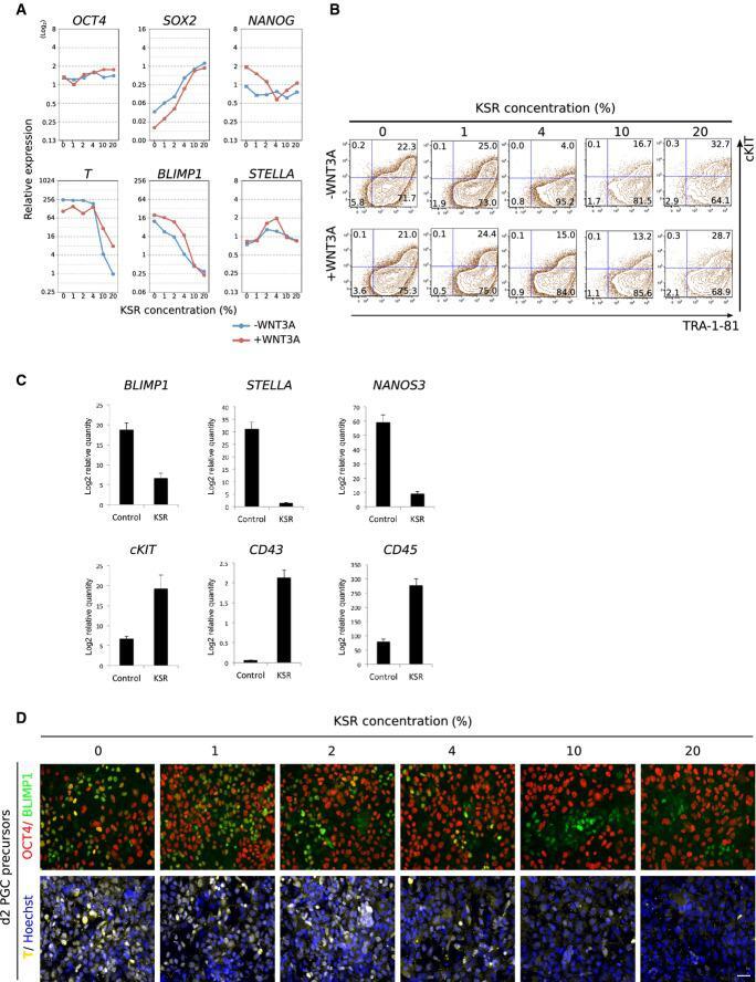
- Figure 4 Effects of KSR and WNT3A on PGC-precursor and PGCLC induction Effects of KSR and WNT3A during PGC-precursor induction on the expression of selected pluripotency, PGC, and a mesodermal gene of d2 cultures. The value for iPSCs is set as 1, and values are on log 2 scale. Data are presented as means +- SD ( n = 3). Effects of KSR and WNT3A on the induction of d6 PGCLCs as analyzed by FACS gated for TRA-1-81 and cKIT. Gene expression analysis of selected PGC and hematopoietic markers in d6 PGCLCs that were cultured in 0% (control) or 20% KSR (KSR) condition during PGC-precursor induction. Samples were calibrated with iPSC values, and iPSC values depict 1. Data are presented as means +- SD ( n = 3). Immunofluorescence analysis for OCT4 (red), BLIMP1 (green), and T (yellow) in d2 precursors cultured in increasing KSR concentrations. Nuclei were stained with Hoechst (blue). Scale bar: 100 mum.
- Submitted by
- Invitrogen Antibodies (provider)
- Main image

- Experimental details
- Figure 5 Transcriptomics profiles during PGC-precursor and PGCLC induction Unsupervised hierarchical clustering of HuES6 ESCs, 383.2 iPSCs, d2 PGC-precursor cultures, and FACS-sorted PGCLCs. Principal component analysis of HuES6 ESCs, 393.2 iPSCs, d2 PGC-precursor cultures, and FACS-sorted PGCLCs. Scatter plots of global gene expression microarrays comparing d2 PGC-precursor cultures, or d4 or d6 FACS-sorted PGCLCs with iPSCs. Venn diagrams of the transcriptomics meta-analysis with the intersections of the DEGs of mouse in vitro PGCLCs, human in vivo PGCs, and human in vitro PGC-like cells in relation to their respective pluripotent counterparts in their respective platforms. Heatmap of pluripotent, germ cell, mesodermal, and chromatin-related markers in mouse (left) and human (right) transcriptomics samples ( Tfap2c is not targeted in the Illumina MouseRef-8 v2). The color bars codify the gene expression in log 2 scale. Red corresponds to high gene expression. Blimp1 +/ Stella + PGCLCs are denoted ""B+S+"", and Blimp1 + /Stella - PGCLCs are denoted ""B+S-"". Effect of knockdown of BLIMP1 and PRDM14 on PGCLC induction. Knockdown of BLIMP1 (F) and PRDM14 (G) was done using the lentiviral system with two individual shRNA vectors for each gene. The knockdown efficiencies were assessed by qPCR (left panel, each). The induction of d4 PGCLCs analyzed by FACS, gated for TRA-1-81 and cKIT (right panel, each). Data are presented as means +- SD ( n = 3).