Explore
 Validate
 Learn
Learn Immunocytochemistry
Antibody data
- Antibody Data
- Antigen structure
- References [11]
- Comments [0]
- Validations
- Immunocytochemistry [6]
- Immunohistochemistry [2]
- Flow cytometry [1]
- Other assay [10]
Submit
Validation data
Reference
Comment
Report error
- Product number
- MA5-12944 - Provider product page

- Provider
- Invitrogen Antibodies
- Product name
- c-Kit Monoclonal Antibody (K45)
- Antibody type
- Monoclonal
- Antigen
- Recombinant protein fragment
- Description
- MA5-12944 targets CD117 in IF/ICC and IHC (P, F) applications and shows reactivity with Human samples. The MA5-12944 immunogen is recombinant human extracellular c-kit fragment.
- Reactivity
- Human
- Host
- Mouse
- Isotype
- IgG
- Antibody clone number
- K45
- Vial size
- 500 μL
- Concentration
- 0.2 mg/mL
- Storage
- 4°C
Submitted references Human Cardiac Progenitor Cells Enhance Exosome Release and Promote Angiogenesis Under Physoxia.
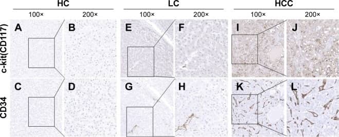
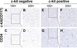
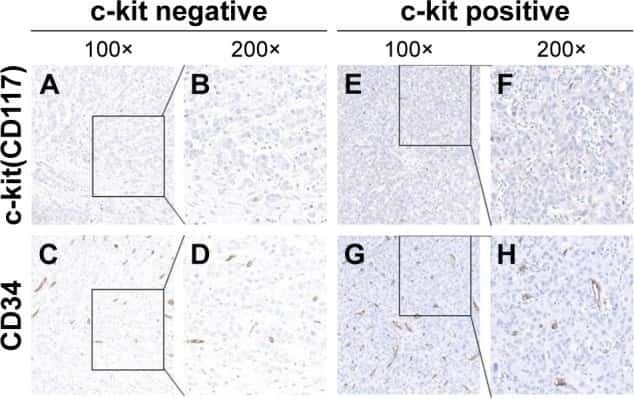
Overexpression of c-kit(CD117), relevant with microvessel density, is an independent survival prognostic factor for patients with HBV-related hepatocellular carcinoma.
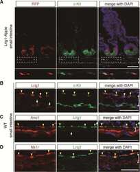
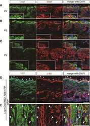
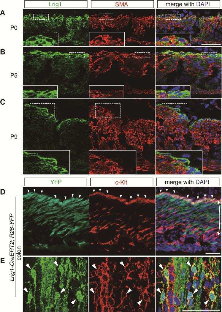
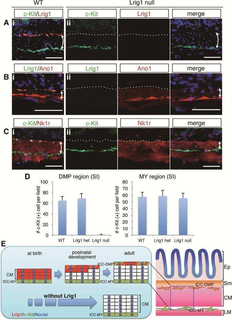
LRIG1 Regulates Ontogeny of Smooth Muscle-Derived Subsets of Interstitial Cells of Cajal in Mice.
Loss of Lrig1 leads to expansion of Brunner glands followed by duodenal adenomas with gastric metaplasia.
Wharton's jelly-derived mesenchymal stem cells promote myocardial regeneration and cardiac repair after miniswine acute myocardial infarction.
Generation of functional blood vessels from a single c-kit+ adult vascular endothelial stem cell.
Septal interstitial cells of Cajal conduct pacemaker activity to excite muscle bundles in human jejunum.
The mechanism and spread of pacemaker activity through myenteric interstitial cells of Cajal in human small intestine.
Evidence for PDGFRA, PDGFRB and KIT deregulation in an NSCLC patient.
Tumor necrosis factor-alpha-converting enzyme controls surface expression of c-Kit and survival of embryonic stem cell-derived mast cells.
Human adult craniofacial muscle-derived cells: neural-cell adhesion-molecule (NCAM; CD56)-expressing cells appear to contain multipotential stem cells.
Dougherty JA, Patel N, Kumar N, Rao SG, Angelos MG, Singh H, Cai C, Khan M
Frontiers in cell and developmental biology 2020;8:130
Frontiers in cell and developmental biology 2020;8:130
Overexpression of c-kit(CD117), relevant with microvessel density, is an independent survival prognostic factor for patients with HBV-related hepatocellular carcinoma.
Yan W, Zhu Z, Pan F, Huang A, Dai GH
OncoTargets and therapy 2018;11:1285-1292
OncoTargets and therapy 2018;11:1285-1292
LRIG1 Regulates Ontogeny of Smooth Muscle-Derived Subsets of Interstitial Cells of Cajal in Mice.
Kondo J, Powell AE, Wang Y, Musser MA, Southard-Smith EM, Franklin JL, Coffey RJ
Gastroenterology 2015 Aug;149(2):407-19.e8
Gastroenterology 2015 Aug;149(2):407-19.e8
Loss of Lrig1 leads to expansion of Brunner glands followed by duodenal adenomas with gastric metaplasia.
Wang Y, Shi C, Lu Y, Poulin EJ, Franklin JL, Coffey RJ
The American journal of pathology 2015 Apr;185(4):1123-34
The American journal of pathology 2015 Apr;185(4):1123-34
Wharton's jelly-derived mesenchymal stem cells promote myocardial regeneration and cardiac repair after miniswine acute myocardial infarction.
Zhang W, Liu XC, Yang L, Zhu DL, Zhang YD, Chen Y, Zhang HY
Coronary artery disease 2013 Nov;24(7):549-58
Coronary artery disease 2013 Nov;24(7):549-58
Generation of functional blood vessels from a single c-kit+ adult vascular endothelial stem cell.
Fang S, Wei J, Pentinmikko N, Leinonen H, Salven P
PLoS biology 2012;10(10):e1001407
PLoS biology 2012;10(10):e1001407
Septal interstitial cells of Cajal conduct pacemaker activity to excite muscle bundles in human jejunum.
Lee HT, Hennig GW, Fleming NW, Keef KD, Spencer NJ, Ward SM, Sanders KM, Smith TK
Gastroenterology 2007 Sep;133(3):907-17
Gastroenterology 2007 Sep;133(3):907-17
The mechanism and spread of pacemaker activity through myenteric interstitial cells of Cajal in human small intestine.
Lee HT, Hennig GW, Fleming NW, Keef KD, Spencer NJ, Ward SM, Sanders KM, Smith TK
Gastroenterology 2007 May;132(5):1852-65
Gastroenterology 2007 May;132(5):1852-65
Evidence for PDGFRA, PDGFRB and KIT deregulation in an NSCLC patient.
Negri T, Casieri P, Miselli F, Orsenigo M, Piacenza C, Stacchiotti S, Bidoli P, Casali PG, Pierotti MA, Tamborini E, Pilotti S
British journal of cancer 2007 Jan 15;96(1):180-1
British journal of cancer 2007 Jan 15;96(1):180-1
Tumor necrosis factor-alpha-converting enzyme controls surface expression of c-Kit and survival of embryonic stem cell-derived mast cells.
Cruz AC, Frank BT, Edwards ST, Dazin PF, Peschon JJ, Fang KC
The Journal of biological chemistry 2004 Feb 13;279(7):5612-20
The Journal of biological chemistry 2004 Feb 13;279(7):5612-20
Human adult craniofacial muscle-derived cells: neural-cell adhesion-molecule (NCAM; CD56)-expressing cells appear to contain multipotential stem cells.
Sinanan AC, Hunt NP, Lewis MP
Biotechnology and applied biochemistry 2004 Aug;40(Pt 1):25-34
Biotechnology and applied biochemistry 2004 Aug;40(Pt 1):25-34
No comments: Submit comment
Supportive validation
- Submitted by
- Invitrogen Antibodies (provider)
- Main image
- Experimental details
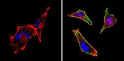
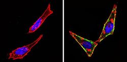
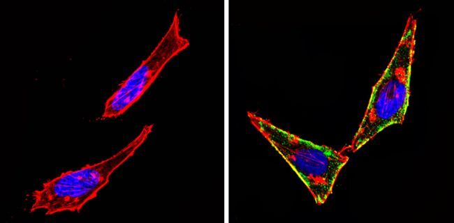
- Immunofluorescent analysis of CD117 (green) showing staining in the cytoplasm and membrane of Hela cells (right) compared to a negative control without primary antibody (left). Formalin-fixed cells were permeabilized with 0.1% Triton X-100 in TBS for 5-10 minutes and blocked with 3% BSA-PBS for 30 minutes at room temperature. Cells were probed with a CD117 monoclonal antibody (Product # MA5-12944) in 3% BSA-PBS at a dilution of 1:50 and incubated overnight at 4 ºC in a humidified chamber. Cells were washed with PBST and incubated with a DyLight-conjugated secondary antibody in PBS at room temperature in the dark. F-actin (red) was stained with a fluorescent red phalloidin and nuclei (blue) were stained with Hoechst or DAPI. Images were taken at a magnification of 60x.

- Submitted by
- Invitrogen Antibodies (provider)
- Main image
- Experimental details
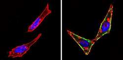
- Immunofluorescent analysis of CD117 (green) showing staining in the cytoplasm and membrane of U-87 MG cells (right) compared to a negative control without primary antibody (left). Formalin-fixed cells were permeabilized with 0.1% Triton X-100 in TBS for 5-10 minutes and blocked with 3% BSA-PBS for 30 minutes at room temperature. Cells were probed with a CD117 monoclonal antibody (Product # MA5-12944) in 3% BSA-PBS at a dilution of 1:50 and incubated overnight at 4 ºC in a humidified chamber. Cells were washed with PBST and incubated with a DyLight-conjugated secondary antibody in PBS at room temperature in the dark. F-actin (red) was stained with a fluorescent red phalloidin and nuclei (blue) were stained with Hoechst or DAPI. Images were taken at a magnification of 60x.

- Submitted by
- Invitrogen Antibodies (provider)
- Main image
- Experimental details
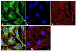
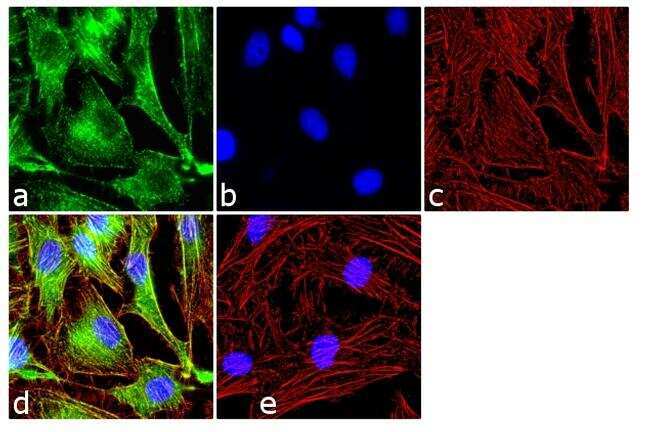
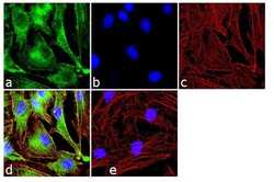
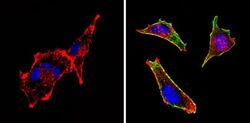
- Immunofluorescence analysis of c-Kit/CD117 was done on 70% confluent log phase HeLa cells. The cells were fixed with 4% paraformaldehyde for 10 minutes, permeabilized with 0.1% Triton™ X-100 for 10 minutes, and blocked with 1% BSA for 1 hour at room temperature. The cells were labeled c-Kit/CD117 (K45) Mouse Monoclonal Antibody (Product # MA5-12944) at 2 µg/mL in 0.1% BSA and incubated for 3 hours at room temperature and then labeled with Goat anti-Mouse IgG (H+L) Superclonal™ Secondary Antibody, Alexa Fluor® 488 conjugate (Product # A28175) at a dilution of 1:2000 for 45 minutes at room temperature (Panel a: green). Nuclei (Panel b: blue) were stained with SlowFade® Gold Antifade Mountant with DAPI (Product # S36938). F-actin (Panel c: red) was stained with Alexa Fluor® 555 Rhodamine Phalloidin (Product # R415, 1:300). Panel d is a merged image showing cytoplasmic and membranous localization. Panel e is a no primary antibody control. The images were captured at 60X magnification.
- Submitted by
- Invitrogen Antibodies (provider)
- Main image
- Experimental details
- Immunofluorescence analysis of c-Kit/CD117 was done on 70% confluent log phase HeLa cells. The cells were fixed with 4% paraformaldehyde for 10 minutes, permeabilized with 0.1% Triton™ X-100 for 10 minutes, and blocked with 1% BSA for 1 hour at room temperature. The cells were labeled c-Kit/CD117 (K45) Mouse Monoclonal Antibody (Product # MA5-12944) at 2 µg/mL in 0.1% BSA and incubated for 3 hours at room temperature and then labeled with Goat anti-Mouse IgG (H+L) Superclonal™ Secondary Antibody, Alexa Fluor® 488 conjugate (Product # A28175) at a dilution of 1:2000 for 45 minutes at room temperature (Panel a: green). Nuclei (Panel b: blue) were stained with SlowFade® Gold Antifade Mountant with DAPI (Product # S36938). F-actin (Panel c: red) was stained with Alexa Fluor® 555 Rhodamine Phalloidin (Product # R415, 1:300). Panel d is a merged image showing cytoplasmic and membranous localization. Panel e is a no primary antibody control. The images were captured at 60X magnification.

- Submitted by
- Invitrogen Antibodies (provider)
- Main image
- Experimental details
- Immunofluorescent analysis of CD117 (green) showing staining in the cytoplasm and membrane of Hela cells (right) compared to a negative control without primary antibody (left). Formalin-fixed cells were permeabilized with 0.1% Triton X-100 in TBS for 5-10 minutes and blocked with 3% BSA-PBS for 30 minutes at room temperature. Cells were probed with a CD117 monoclonal antibody (Product # MA5-12944) in 3% BSA-PBS at a dilution of 1:50 and incubated overnight at 4 ºC in a humidified chamber. Cells were washed with PBST and incubated with a DyLight-conjugated secondary antibody in PBS at room temperature in the dark. F-actin (red) was stained with a fluorescent red phalloidin and nuclei (blue) were stained with Hoechst or DAPI. Images were taken at a magnification of 60x.
- Submitted by
- Invitrogen Antibodies (provider)
- Main image
- Experimental details
- Immunofluorescent analysis of CD117 (green) showing staining in the cytoplasm and membrane of U-87 MG cells (right) compared to a negative control without primary antibody (left). Formalin-fixed cells were permeabilized with 0.1% Triton X-100 in TBS for 5-10 minutes and blocked with 3% BSA-PBS for 30 minutes at room temperature. Cells were probed with a CD117 monoclonal antibody (Product # MA5-12944) in 3% BSA-PBS at a dilution of 1:50 and incubated overnight at 4 ºC in a humidified chamber. Cells were washed with PBST and incubated with a DyLight-conjugated secondary antibody in PBS at room temperature in the dark. F-actin (red) was stained with a fluorescent red phalloidin and nuclei (blue) were stained with Hoechst or DAPI. Images were taken at a magnification of 60x.

Supportive validation
- Submitted by
- Invitrogen Antibodies (provider)
- Main image
- Experimental details
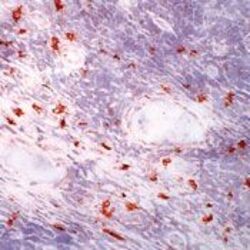
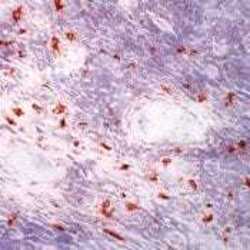
- Frozen human tonsil section stained with CD117 antibody using UltraVision LP and AEC chromogen. Note staining of mast cells.

- Submitted by
- Invitrogen Antibodies (provider)
- Main image
- Experimental details
- Frozen human tonsil section stained with CD117 antibody using UltraVision LP and AEC chromogen. Note staining of mast cells.

Supportive validation
- Submitted by
- Invitrogen Antibodies (provider)
- Main image
- Experimental details
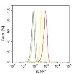
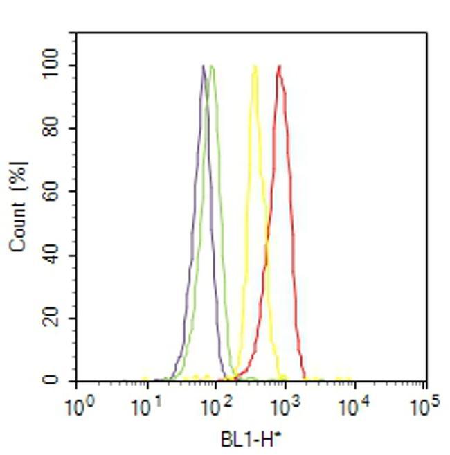
- Flow cytometry analysis of c-Kit / CD117 was done on A549 cells. Cells were fixed with 70% ethanol for 10 minutes, permeabilized with 0.25% Triton™ X-100 for 20 minutes, and blocked with 5% BSA for 30 minutes at room temperature. Cells were labeled with c-Kit / CD117 Mouse Monoclonal Antibody (MA512944, red histogram) or with mouse isotype control (yellow histogram) at 3-5 ug/million cells in 2.5% BSA. After incubation at room temperature for 2 hours, the cells were labeled with Alexa Fluor® 488 Rabbit Anti-Mouse Secondary Antibody (A11059) at a dilution of 1:400 for 30 minutes at room temperature. The representative 10,000 cells were acquired and analyzed for each sample using an Attune® Acoustic Focusing Cytometer. The purple histogram represents unstained control cells and the green histogram represents no-primary-antibody control.
Supportive validation
- Submitted by
- Invitrogen Antibodies (provider)
- Main image
- Experimental details
- NULL
- Submitted by
- Invitrogen Antibodies (provider)
- Main image
- Experimental details
- NULL
- Submitted by
- Invitrogen Antibodies (provider)
- Main image
- Experimental details
- NULL
- Submitted by
- Invitrogen Antibodies (provider)
- Main image
- Experimental details
- NULL
- Submitted by
- Invitrogen Antibodies (provider)
- Main image

- Experimental details
- NULL
- Submitted by
- Invitrogen Antibodies (provider)
- Main image
- Experimental details
- NULL
- Submitted by
- Invitrogen Antibodies (provider)
- Main image
- Experimental details
- NULL
- Submitted by
- Invitrogen Antibodies (provider)
- Main image

- Experimental details
- NULL
- Submitted by
- Invitrogen Antibodies (provider)
- Main image
- Experimental details
- NULL
- Submitted by
- Invitrogen Antibodies (provider)
- Main image

- Experimental details
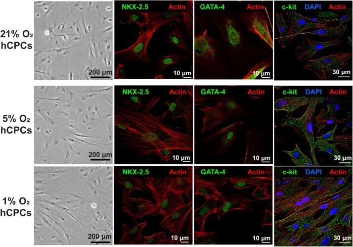
- FIGURE 1 hCPCs morphology and cardiac gene expression under normoxic and hypoxic microenvironments. Human CPCs were cultured under 21, 5, and 1% O 2 for 48 h. DIC imaging shows the typical morphology of cells, which is unchanged under hypoxia. Immunofluorescent staining for cardiac lineage markers NKX-2.5 (green, nuclear), GATA-4 (green, nuclear), and c-kit (green) showed their expression was maintained under all oxygen conditions. Nuclei are stained blue and F-actin is stained red.