Explore
 Validate
 Learn
Learn Western blot
Antibody data
- Antibody Data
- Antigen structure
- References [0]
- Comments [0]
- Validations
- Western blot [2]
- Immunocytochemistry [2]
- Immunohistochemistry [2]
Submit
Validation data
Reference
Comment
Report error
- Product number
- PA5-29233 - Provider product page

- Provider
- Invitrogen Antibodies
- Product name
- Cyclin B2 Polyclonal Antibody
- Antibody type
- Polyclonal
- Antigen
- Recombinant protein fragment
- Description
- Recommended positive controls: H1299.
- Concentration
- 1 mg/mL
No comments: Submit comment
Supportive validation
- Submitted by
- Invitrogen Antibodies (provider)
- Main image

- Experimental details
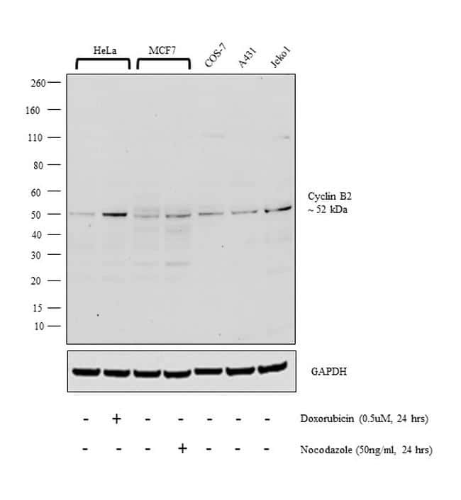
- Western blot analysis was performed on whole cell extract (30 µg lysate) of HeLa (Lane 1), HeLa treated with Doxorubicin (0.5uM, 24 hrs) (Lane 2), MCF7 (Lane 3), MCF7 treated with Nocodazole (50ng/ml, 24 hrs) (Lane 4), COS-7 (Lane 5), A431 (Lane 6), and Jeko1 (Lane 7). The blot was probed with Anti-Cyclin B2 Polyclonal Antibody (Product # PA5-29233, 1:2000 dilution) and detected by chemiluminescence using Goat anti-Rabbit IgG (H+L) Superclonal™ Secondary Antibody, HRP conjugate (Product # A27036, 0.25 µg/ml, 1:4000 dilution). A 52 kDa band corresponding to Cyclin B2 was observed in all cell lines tested and was enhanced upon treatment.
- Submitted by
- Invitrogen Antibodies (provider)
- Main image
- Experimental details
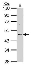
- Western Blot using Cyclin B2 Polyclonal Antibody (Product # PA5-29233). Sample (30 µg of whole cell lysate). Lane A: H1299. 10% SDS PAGE. Cyclin B2 Polyclonal Antibody (Product # PA5-29233) diluted at 1:1,000.

Supportive validation
- Submitted by
- Invitrogen Antibodies (provider)
- Main image
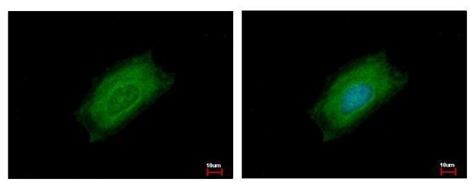
- Experimental details
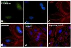
- Immunofluorescence analysis of Cyclin B2 was performed using 70% confluent log phase HeLa cells treated with Camptothecin (100nM, 16 hrs). The cells were fixed with 4% paraformaldehyde for 10 minutes, permeabilized with 0.1% Triton™ X-100 for 15 minutes, and blocked with 1% BSA for 1 hour at room temperature. The cells were labeled with Cyclin B2 Polyclonal Antibody (Product # PA5-29233) at 1:200 dilution in 0.1% BSA, incubated at 4 degree Celsius overnight and then labeled with Goat anti-Rabbit IgG (H+L) Superclonal™ Secondary Antibody, Alexa Fluor® 488 conjugate (Product # A27034) at a dilution of 1:2000 for 45 minutes at room temperature (Panel a: green). Nuclei (Panel b: blue) were stained with ProLong™ Diamond Antifade Mountant with DAPI (Product # P36962). F-actin (Panel c: red) was stained with Rhodamine Phalloidin (Product # R415, 1:300). Panel d represents the merged image showing accumulation of Cyclin A2 in the nucleus upon treatment. Panel e represents the control cells showing cytoplasmic staining. Panel f represents control cells with no primary antibody to assess background. The images were captured at 60X magnification.

- Submitted by
- Invitrogen Antibodies (provider)
- Main image
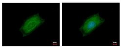
- Experimental details
- Cyclin B2 Polyclonal Antibody [N1C3-2] detects CCNB2 protein at cytoplasm by immunofluorescent analysis. Sample: HeLa cells were fixed in ice-cold MeOH for 5 min. Green: CCNB2 protein stained by Cyclin B2 Polyclonal Antibody [N1C3-2] (Product # PA5-29233) diluted at 1:500. Blue: Hoechst 33343 staining.
Supportive validation
- Submitted by
- Invitrogen Antibodies (provider)
- Main image

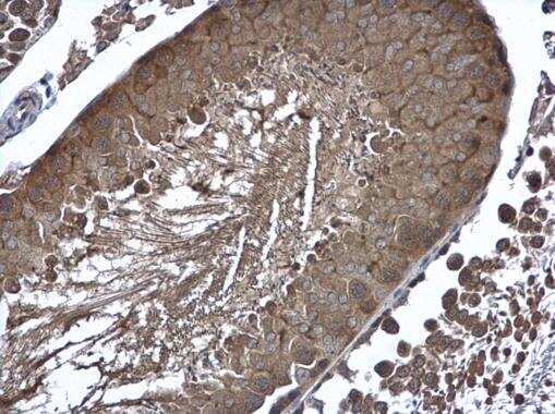
- Experimental details
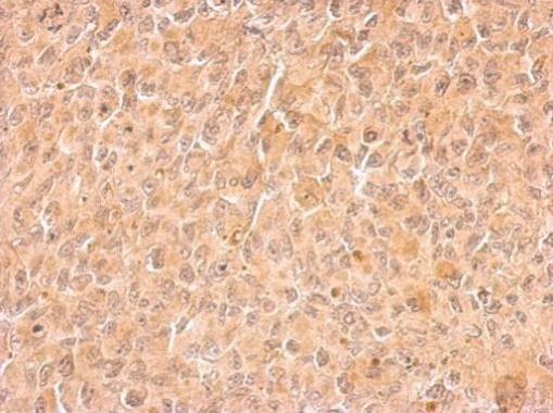
- Immunohistochemistry (Paraffin) analysis of Cyclin B2 was performed in paraffin-embedded rat testis tissue using Cyclin B2 Polyclonal Antibody (Product # PA5-29233) at a dilution of 1:500.
- Submitted by
- Invitrogen Antibodies (provider)
- Main image
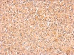
- Experimental details
- Cyclin B2 Polyclonal Antibody detects CCNB2 protein at cytosol on H1299 xenograft by immunohistochemical analysis. Sample: Paraffin-embedded H1299 xenograft. Cyclin B2 Polyclonal Antibody (Product # PA5-29233) dilution: 1:500. Antigen Retrieval: EDTA based buffer, pH 8.0, 15 min.