Explore
 Validate
 Learn
Learn Western blot
 Immunocytochemistry
Antibody data
- Antibody Data
- Antigen structure
- References [0]
- Comments [0]
- Validations
- Immunocytochemistry [2]
- Immunohistochemistry [1]
Submit
Validation data
Reference
Comment
Report error
- Product number
- PA5-28050 - Provider product page

- Provider
- Invitrogen Antibodies
- Product name
- BCKDK Polyclonal Antibody
- Antibody type
- Polyclonal
- Antigen
- Recombinant full-length protein
- Description
- Recommended positive controls: MCF-7, MCF-7 membrane extract, Mouse brain, rat brain, rat kidney. Predicted reactivity: Mouse (99%), Rat (100%), Zebrafish (82%), Xenopus laevis (87%), Rhesus Monkey (98%), Bovine (98%). Store product as a concentrated solution. Centrifuge briefly prior to opening the vial.
- Reactivity
- Human, Mouse, Rat
- Host
- Rabbit
- Isotype
- IgG
- Vial size
- 100 μL
- Concentration
- 0.35 mg/mL
- Storage
- Store at 4°C short term. For long term storage, store at -20°C, avoiding freeze/thaw cycles.
No comments: Submit comment
Supportive validation
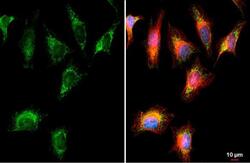
- Submitted by
- Invitrogen Antibodies (provider)
- Main image

- Experimental details
- BCKDK Polyclonal Antibody detects BCKDK protein at mitochondria by immunofluorescent analysis. Sample: HeLa cells were fixed in 4% paraformaldehyde at RT for 15 min. Green: BCKDK stained by BCKDK Polyclonal Antibody (Product # PA5-28050) diluted at 1:500. Red: alpha Tubulin, a cytoskeleton marker, stained by alpha Tubulin Polyclonal Antibody [GT114] (Product # MA5-31466) diluted at 1:1,000. Blue: Fluoroshield with DAPI .

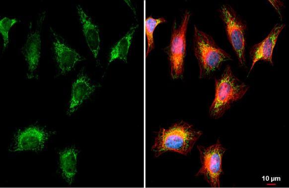
- Submitted by
- Invitrogen Antibodies (provider)
- Main image
- Experimental details
- BCKDK Polyclonal Antibody detects BCKDK protein at mitochondria by immunofluorescent analysis. Sample: HeLa cells were fixed in 4% paraformaldehyde at RT for 15 min. Green: BCKDK stained by BCKDK Polyclonal Antibody (Product # PA5-28050) diluted at 1:500. Red: alpha Tubulin, a cytoskeleton marker, stained by alpha Tubulin Polyclonal Antibody [GT114] (Product # MA5-31466) diluted at 1:1,000. Blue: Fluoroshield with DAPI .
Supportive validation
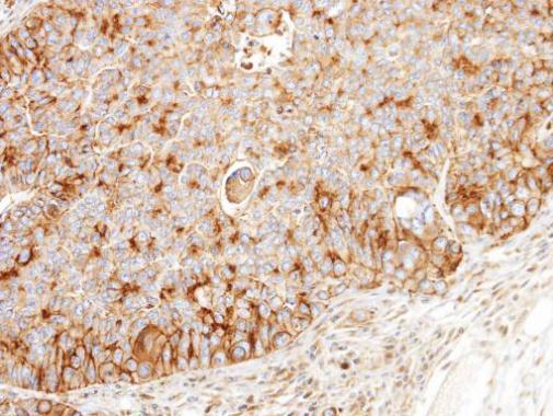
- Submitted by
- Invitrogen Antibodies (provider)
- Main image
- Experimental details
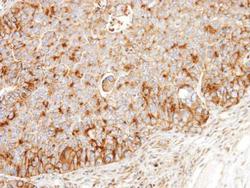
- Immunohistochemical analysis of paraffin-embedded SW480 xenograft, using BCKDK (Product # PA5-28050) antibody at 1:100 dilution. Antigen Retrieval: EDTA based buffer, pH 8.0, 15 min.