 Explore
 Validate
 Learn
Learn Western blot
Antibody data
- Antibody Data
- Antigen structure
- References [0]
- Comments [0]
- Validations
- Western blot [4]
- Immunocytochemistry [2]
- Immunohistochemistry [1]
Submit
Validation data
Reference
Comment
Report error
- Product number
- PA5-21877 - Provider product page

- Provider
- Invitrogen Antibodies
- Product name
- NLK Polyclonal Antibody
- Antibody type
- Polyclonal
- Antigen
- Recombinant protein fragment
- Description
- Recommended positive controls: NLK-transfected 293T, A431, H1299, HeLa, mouse brain.
- Concentration
- 1 mg/mL
No comments: Submit comment
Supportive validation
- Submitted by
- Invitrogen Antibodies (provider)
- Main image
- Experimental details
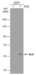
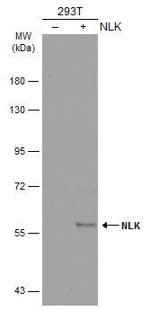
- Western Blot analysis of NLK was performed by separating 30 µg of non-transfected (–) and transfected (+) 293T whole cell extracts by 10% SDS-PAGE. Proteins were transferred to a membrane and probed with a NLK Polyclonal Antibody (Product # PA5-21877) at a dilution of 1:500. The HRP-conjugated anti-rabbit IgG antibody was used to detect the primary antibody.
- Submitted by
- Invitrogen Antibodies (provider)
- Main image

- Experimental details
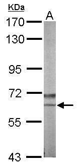
- Western Blot using NLK Polyclonal Antibody (Product # PA5-21877). Sample (50 µg of whole cell lysate). Lane A: Mouse brain. 7.5% SDS PAGE. NLK Polyclonal Antibody (Product # PA5-21877) diluted at 1:1,000. The HRP-conjugated anti-rabbit IgG antibody was used to detect the primary antibody.
- Submitted by
- Invitrogen Antibodies (provider)
- Main image

- Experimental details
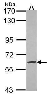
- Western Blot using NLK Polyclonal Antibody (Product # PA5-21877). Sample (30 µg of whole cell lysate). Lane A: HeLa. 7.5% SDS PAGE. NLK antibody. NLK Polyclonal Antibody (Product # PA5-21877) diluted at 1:1,000. The HRP-conjugated anti-rabbit IgG antibody was used to detect the primary antibody.
- Submitted by
- Invitrogen Antibodies (provider)
- Main image

- Experimental details
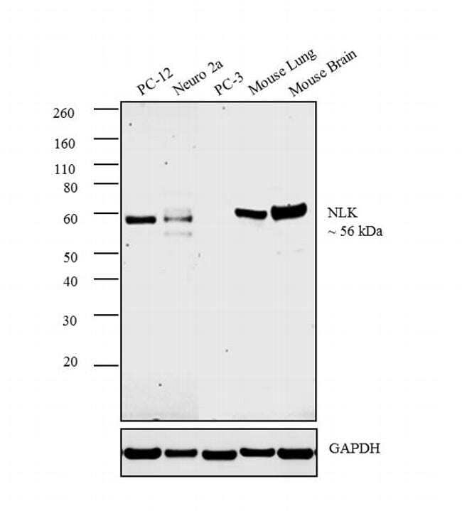
- Western blot analysis was performed on whole cell extracts (30 µg lysate) of PC-12 (Lane 1), Neuro 2a (Lane 2), PC-3 (Lane 3) and tissue extracts of Mouse Lung (Lane 4) and Mouse brain (Lane 5). The blot was probed with Anti- NLK Polyclonal Antibody (Product # PA5-21877, 1:1000 dilution) and detected by chemiluminescence using Goat anti-Rabbit IgG (H+L) Superclonal™ Secondary Antibody, HRP conjugate (Product # A28177, 0.25 µg/mL, 1:4000 dilution). A 56 kDa band corresponding to NLK was observed across all the cell lines and tissues tested except PC-3 cell line.
Supportive validation
- Submitted by
- Invitrogen Antibodies (provider)
- Main image
- Experimental details
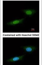
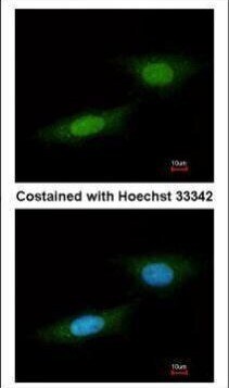
- Immunofluorescent analysis of NLK in paraformaldehyde-fixed HeLa cells using a NLK polyclonal antibody (Product # PA5-21877) at a 1:100 dilution.
- Submitted by
- Invitrogen Antibodies (provider)
- Main image

- Experimental details
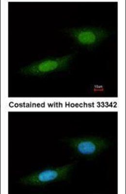
- Immunofluorescence analysis of paraformaldehyde-fixed HeLa, using NLK (Product # PA5-21877) antibody at 1:100 dilution.
Supportive validation
- Submitted by
- Invitrogen Antibodies (provider)
- Main image
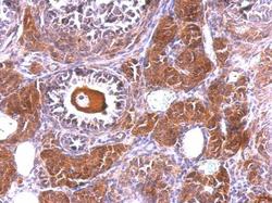
- Experimental details
- NLK Polyclonal Antibody detects NLK protein at cytosol and nucleus on mouse ovary by immunohistochemical analysis. Sample: Paraffin-embedded mouse ovary. NLK Polyclonal Antibody (Product # PA5-21877) dilution: 1:500. Antigen Retrieval: EDTA based buffer, pH 8.0, 15 min.
