Explore
 Validate
 Learn
Learn Western blot
 Immunocytochemistry
 Immunohistochemistry
Antibody data
- Antibody Data
- Antigen structure
- References [1]
- Comments [0]
- Validations
- Western blot [1]
- Immunocytochemistry [1]
Submit
Validation data
Reference
Comment
Report error
- Product number
- HPA004689 - Provider product page

- Provider
- Atlas Antibodies
- Proper citation
- Atlas Antibodies Cat#HPA004689, RRID:AB_1078194
- Product name
- Anti-ARHGAP1
- Antibody type
- Polyclonal
- Description
- Polyclonal Antibody against Human ARHGAP1, Gene description: Rho GTPase activating protein 1, Alternative Gene Names: CDC42GAP, p50rhoGAP, RhoGAP, Validated applications: ICC, IHC, WB, Uniprot ID: Q07960, Storage: Store at +4°C for short term storage. Long time storage is recommended at -20°C.
- Reactivity
- Human, Mouse, Rat
- Host
- Rabbit
- Conjugate
- Unconjugated
- Isotype
- IgG
- Vial size
- 100 µl
- Concentration
- 0.05 mg/ml
- Storage
- Store at +4°C for short term storage. Long time storage is recommended at -20°C.
- Handling
- The antibody solution should be gently mixed before use.
Submitted references Lysophosphatidic acid and sphingosine-1-phosphate promote morphogenesis and block invasion of prostate cancer cells in three-dimensional organotypic models
Härmä V, Knuuttila M, Virtanen J, Mirtti T, Kohonen P, Kovanen P, Happonen A, Kaewphan S, Ahonen I, Kallioniemi O, Grafström R, Lötjönen J, Nees M
Oncogene 2011;31(16):2075-2089
Oncogene 2011;31(16):2075-2089
No comments: Submit comment
Enhanced validation
- Submitted by
- Atlas Antibodies (provider)
- Enhanced method
- Genetic validation
- Main image
- Experimental details
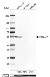
- Western blot analysis in U-138MG cells transfected with control siRNA, target specific siRNA probe #1 and #2, using Anti-ARHGAP1 antibody. Remaining relative intensity is presented. Loading control: Anti-GAPDH.
- Sample type
- Human
- Protocol
- Protocol

Supportive validation
- Submitted by
- Atlas Antibodies (provider)
- Main image
- Experimental details
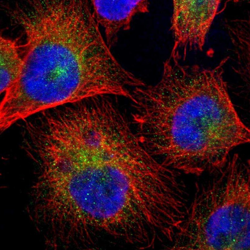
- Immunofluorescent staining of human cell line U-251 MG shows localization to vesicles.
- Sample type
- Human