Explore
 Validate
 Learn
Learn Western blot
 Immunoprecipitation
Antibody data
- Antibody Data
- Antigen structure
- References [0]
- Comments [0]
- Validations
- Western blot [2]
- Immunocytochemistry [1]
- Immunohistochemistry [2]
- Other assay [1]
Submit
Validation data
Reference
Comment
Report error
- Product number
- AGC-002-200UL - Provider product page

- Provider
- Invitrogen Antibodies
- Product name
- NMDAR2A (GluN2A) (extracellular) Polyclonal Antibody
- Antibody type
- Polyclonal
- Antigen
- Other
- Reactivity
- Human, Mouse, Rat
- Host
- Rabbit
- Isotype
- IgG
- Vial size
- 200 µL
- Concentration
- 0.55 mg/mL
- Storage
- -20° C, Avoid Freeze/Thaw Cycles
No comments: Submit comment
Supportive validation
- Submitted by
- Invitrogen Antibodies (provider)
- Main image
- Experimental details
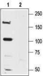
- Western blot analysisof rat brain lysates: - 1. Anti-NMDAR2A (GluN2A) (extracellular) Antibody (#AGC-002), (1:600). 2. Anti-NMDAR2A (GluN2A) (extracellular) Antibody , preincubated with NMDAR2A/GluN2A (extracellular) Blocking Peptide (#BLP-GC002).

- Submitted by
- Invitrogen Antibodies (provider)
- Main image

- Experimental details
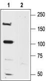
- Western blot analysisof rat brain lysates: - 1. Anti-NMDAR2A (GluN2A) (extracellular) Antibody (#AGC-002), (1:600). 2. Anti-NMDAR2A (GluN2A) (extracellular) Antibody , preincubated with NMDAR2A/GluN2A (extracellular) Blocking Peptide (#BLP-GC002).
Supportive validation
- Submitted by
- Invitrogen Antibodies (provider)
- Main image
- Experimental details
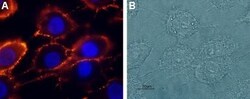
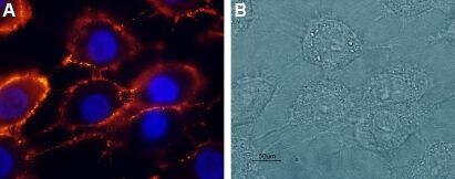
- Expression of NR2A in rat C6 glioma cells - Cell surface detection of NR2A in live intact rat C6 glioma cells. A. Cells werestained with Anti-NMDAR2A (GluN2A) (extracellular) Antibody (#AGC-002), (1:100), followed by goat Anti-rabbit-AlexaFluor-555 secondary Antibody (red). Cell nuclei were stained with the cell permeable dye Hoechst 33342 (blue staining). B. Live view of the same field.
Supportive validation
- Submitted by
- Invitrogen Antibodies (provider)
- Main image
- Experimental details
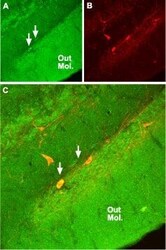
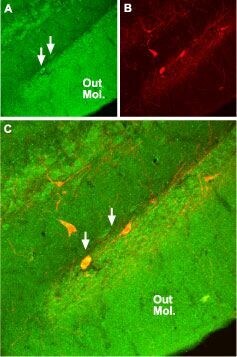
- Expression of NR2A in rat hippocampus - Immunohistochemical staining of rat hippocampaldentate gyrus with Anti-NMDAR2A (GluN2A) (extracellular) Antibody (#AGC-002). A.NMDAR2A (green) appears diffusely in the outer molecular layer of thedentate gyrus (Out Mol.) and in cells along the subgranular layer (arrows). B. Staining of parvalbumin (PV, red) identifies interneurons in thedentate gyrus. C. Confocal merge demonstrates localization of PV in some neurons with NMDAR2A.
- Submitted by
- Invitrogen Antibodies (provider)
- Main image

- Experimental details
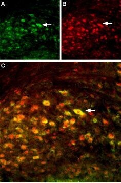
- Multiplex staining of GluN2A and GluN2B in mouse deep cerebellar nucleus - Immunohistochemical staining of perfusion-fixed frozen mouse brain sections using Anti-NMDAR2B (GluN2B) (extracellular)-ATTO Fluor-594 Antibody (#AGC-003-AR), (1:60) and Anti-NMDAR2A (GluN2A) (extracellular) Antibody (#AGC-002), (1:200). A. Sections were incubated with Anti-NMDAR2A (GluN2A) (extracellular) Antibody , followed by goat Anti-rabbit-Alexa-488 (green). B. The same sections were incubated with Anti-NMDAR2B (GluN2B) (extracellular)-ATTO Fluor-594 Antibody (red). C. Merge of A and B demonstrates the ubiquitous colocalization of the GluN2A and GluN2B subunits in cells with neuronal profiles in this nucleus. Arrows point at an example of NR2A and NR2B co-expression.
Supportive validation
- Submitted by
- Invitrogen Antibodies (provider)
- Main image

- Experimental details
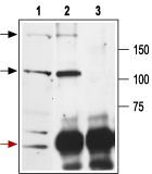
- Immunoprecipitation of rat brain lysates - 1. Cell lysate. 2. Cell lysates + protein A beads + Anti-NMDAR2A (GluN2A) (extracellular) Antibody (#AGC-002).3. Cell lysates + protein A beads + pre-immune rabbit serum.Black arrow indicates the NR2A protein while the red arrow shows the IgG heavy chain. Immunoblot was performed with Anti-NMDAR2A (GluN2A) (extracellular) Antibody .