Explore
 Validate
 Learn
Learn Western blot
 Immunohistochemistry
Antibody data
- Antibody Data
- Antigen structure
- References [1]
- Comments [0]
- Validations
- Immunohistochemistry [1]
Submit
Validation data
Reference
Comment
Report error
- Product number
- PB9960 - Provider product page

- Provider
- Boster Biological Technology
- Product name
- Anti-Glucose Transporter 5 GLUT5/SLC2A5 Antibody Picoband™
- Antibody type
- Polyclonal
- Description
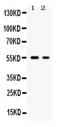
- Polyclonal antibody for Glut5/SLC2A5 detection. Host: Rabbit.Size: 100μg/vial. Tested applications: WB, IHC-P, IHC-F, ICC/IF, FCM. Reactive species: Human. Glut5/SLC2A5 information: Molecular Weight: 54974 MW; Subcellular Localization: Apical cell membrane ; Multi- pass membrane protein . Membrane; Multi-pass membrane protein. Localized on the apical membrane of the small intestine and the proximal tubule of the kidney; Tissue Specificity: Expressed in small intestine, and at much lower levels in kidney, skeletal muscle, and adipose tissue.
- Reactivity
- Human, Rat
- Host
- Rabbit
- Vial size
- 100μg/vial
- Concentration
- Add 0.2ml of distilled water will yield a concentration of 500ug/ml.
- Storage
- At -20°C for one year. After reconstitution, at 4°C for one month. It can also be aliquoted and stored frozen at -20°C for a longer time. Avoid repeated freezing and thawing.
- Handling
- Add 0.2ml of distilled water will yield a concentration of 500ug/ml.
Submitted references High-fructose consumption suppresses insulin signaling pathway accompanied by activation of macrophage and apoptotic markers in rat testis.
Yildirim OG, Guney C, Alcigir ME, Akar F
Reproductive biology 2023 Dec;23(4):100815
Reproductive biology 2023 Dec;23(4):100815
No comments: Submit comment
Supportive validation
- Submitted by
- Boster Biological Technology (provider)
- Main image
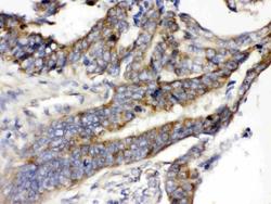
- Experimental details
- IHC analysis of SLC2A5 using anti-SLC2A5 antibody (PB9960). SLC2A5 was detected in paraffin-embedded section of human intestinal cancer tissues. Heat mediated antigen retrieval was performed in citrate buffer (pH6, epitope retrieval solution) for 20 mins. The tissue section was blocked with 10% goat serum. The tissue section was then incubated with 1μg/ml rabbit anti-SLC2A5 Antibody (PB9960) overnight at 4°C. Biotinylated goat anti-rabbit IgG was used as secondary antibody and incubated for 30 minutes at 37°C. The tissue section was developed using Strepavidin-Biotin-Complex (SABC)(Catalog # SA1022) with DAB as the chromogen.
- Additional image