Explore
 Validate
 Learn
Learn Western blot
 ELISA
ELISAAntibody data
- Antibody Data
- Antigen structure
- References [1]
- Comments [0]
- Validations
- ELISA [2]
- Immunohistochemistry [1]
- Other assay [2]
Submit
Validation data
Reference
Comment
Report error
- Product number
- MA5-24530 - Provider product page

- Provider
- Invitrogen Antibodies
- Product name
- ZNF92 Monoclonal Antibody (1F2)
- Antibody type
- Monoclonal
- Antigen
- Recombinant full-length protein
- Description
- Peptide Sequence: FNQSSIFTKH KIIHTEGKSY KCEKCGNAFN QSSNLTARKI IYTGEKPYKY EECDKAFNKF STLITHQIIY TGEKPCKHEC GRAFNKSSNY TKEKLQT
- Reactivity
- Human
- Host
- Mouse
- Isotype
- IgG
- Antibody clone number
- 1F2
- Vial size
- 100 μg
- Concentration
- 1 mg/mL
- Storage
- -20°C, Avoid Freeze/Thaw Cycles
Submitted references ZNF92, an unexplored transcription factor with remarkably distinct breast cancer over-expression associated with prognosis and cell-of-origin.
Kamran M, Bhattacharya U, Omar M, Marchionni L, Ince TA
NPJ breast cancer 2022 Aug 29;8(1):99
NPJ breast cancer 2022 Aug 29;8(1):99
No comments: Submit comment
Supportive validation
- Submitted by
- Invitrogen Antibodies (provider)
- Main image

- Experimental details
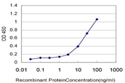
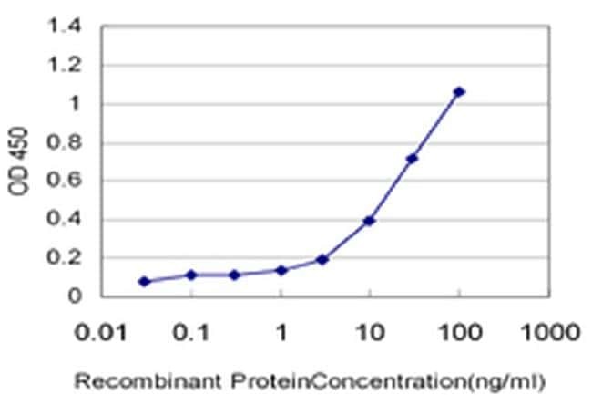
- ELISA analysis using recombinant GST tagged ZNF92 Antibody (Product # MA5-24530) at 0.1 ng/mL as the capture antibody.

- Submitted by
- Invitrogen Antibodies (provider)
- Main image
- Experimental details
- ELISA analysis using recombinant GST tagged ZNF92 Antibody (Product # MA5-24530) at 0.1 ng/mL as the capture antibody.

Supportive validation
- Submitted by
- Invitrogen Antibodies (provider)
- Main image
- Experimental details
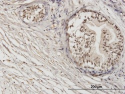
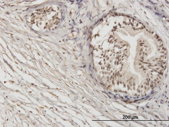
- Immunohistochemical analysis of ZNF92 in formalin-fixed paraffin-embedded human prostate tissue. Samples were probed using a Mouse Monoclonal ZNF92 (1F2) Antibody (Product # MA5-24530) at 1.2 µg/mL.
Supportive validation
- Submitted by
- Invitrogen Antibodies (provider)
- Main image

- Experimental details
- ELISA analysis using recombinant GST tagged ZNF92 Antibody (Product # MA5-24530) at 0.1 ng/mL as the capture antibody.
- Submitted by
- Invitrogen Antibodies (provider)
- Main image

- Experimental details
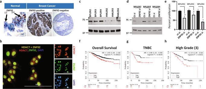
- Fig. 6 ZNF92 expression and function. a Immunohistochemical stain of formalin fixed paraffin embedded sections probed with ZNF92 antibody. The normal breast demonstrates two subgroups of nuclear ZNF92-positive and ZNF92-negative cells (left panel, scale bar = 20 uM). In some breast cancers nearly all tumor cells are ZNF92-positive (middle panel) and other tumors are entirely ZNF92-negative (right panel, scale bar = 100 uM). b Immunofluorescent staining of HDAC7 (red), ZNF92 (green) and nuclear DAPI (blue) in BT20 cells. The merged panel demonstrates the co-expression of HDAC7 and ZNF92 in the same nuclei. The brightness of the entire digital image was increased in the single channel panels to visualize the lower cytoplasmic staining and the individual color channels were adjusted in the merged image for clarity. The uncropped and unprocessed images are provided in Supplementary Fig. 13 . c Western blot analysis of ZNF92 protein expression in matched pairs of BPLER/HMLER 2, 3 and 4. d Western blot analysis of ZNF92 protein after siRNA knock-down of ZNF92 expression (ZNF92 si) in three independent BPLER lines compared to control siRNA (ct si). The uncropped and unprocessed western blot images are provided in Supplementary Fig. 14 . e Bar graphs showing that siRNA knock-down of ZNF92 expression (ZNF92 si) results in statistically significant reduction (*) in cell numbers in all three independent BPLER lines compared to control siRNA (ct si). f - h KM-plot survival analysis of th