 Explore
 Validate
 Learn
Learn Western blot
 Immunoprecipitation
Antibody data
- Antibody Data
- Antigen structure
- References [1]
- Comments [0]
- Validations
- Western blot [2]
- Other assay [1]
Submit
Validation data
Reference
Comment
Report error
- Product number
- PA5-80343 - Provider product page

- Provider
- Invitrogen Antibodies
- Product name
- Apelin Receptor Polyclonal Antibody
- Antibody type
- Polyclonal
- Antigen
- Synthetic peptide
- Description
- This product is preservative free. It is recommended to add sodium azide to avoid contamination (final concentration 0.05%-0.1%). This antibody has specificity for Human APJ Receptor/APLNR.
- Reactivity
- Human
- Host
- Rabbit
- Isotype
- IgG
- Vial size
- 100 µL
- Concentration
- 1 mg/mL
- Storage
- Store at 4°C short term. For long term storage, store at -20°C, avoiding freeze/thaw cycles.
Submitted references Roles for heterodimerization of APJ and B2R in promoting cell proliferation via ERK1/2-eNOS signaling pathway.
Ji B, Shang L, Wang C, Wan L, Cheng B, Chen J
Cellular signalling 2020 Sep;73:109671
Cellular signalling 2020 Sep;73:109671
No comments: Submit comment
Supportive validation
- Submitted by
- Invitrogen Antibodies (provider)
- Main image

- Experimental details
- Knockdown of Apelin receptor was achieved by transfecting K-562 with Apelin receptor specific siRNAs (Silencer® select Product # s1186 and s1188). Western blot analysis (Fig. a) was performed using Whole cell extracts from the Apelin receptor knockdown cells (lane 3), non-targeting scrambled siRNA transfected cells (lane 2) and untransfected cells (lane 1). The blot was probed with Apelin Receptor Polyclonal Antibody (Product # PA5-80343, 0.2 µg/ml) and Goat anti-Rabbit IgG (H+L) Superclonal™ Recombinant Secondary Antibody, HRP (Product # A27036, 1:4000 dilution). Densitometric analysis of this western blot is shown in histogram (Fig. b). Decrease in signal upon siRNA mediated knock down confirms that antibody is specific to Apelin receptor.

- Submitted by
- Invitrogen Antibodies (provider)
- Main image
- Experimental details
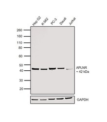
- Western blot was performed using Anti-Apelin Receptor Polyclonal Antibody (Product # PA5-80343) and a 42 kDa band corresponding to Apelin receptor was observed across cell lines tested. Whole cell extracts (20 µg lysate) of Hep G2 (Lane 1), K-562 (Lane 2), PC-3 (Lane 3), Daudi (Lane 4) and Jurkat (Lane 5) were electrophoresed using NuPAGE™ 4-12% Bis-Tris Protein Gel (Product # NP0322BOX). Jurkat expresses APLNR protein at lower levels as compared to Daudi. Resolved proteins were then transferred onto a Nitrocellulose membrane (Product # IB23002) by iBlot® 2 Dry Blotting System (Product # IB21001). The blot was probed with the primary antibody (0.2 µg/ml) and detected by chemiluminescence with Goat anti-Rabbit IgG (H+L) Superclonal™ Recombinant Secondary Antibody, HRP (Product # A27036, 1:4000 dilution) using the iBright FL 1000 (Product # A32752). Chemiluminescent detection was performed using Novex® ECL Chemiluminescent Substrate Reagent Kit (Product # WP20005).

Supportive validation
- Submitted by
- Invitrogen Antibodies (provider)
- Main image

- Experimental details
- Apelin Receptor Immunoprecipitation using: Lane A: 0.5 mg Mouse heart tissue lysate 4 µL with Apelin Receptor Polyclonal Antibody (Product # PA5-80343) and 60 μg of Immunomagnetic beads Protein A/G.Primary antibody: Apelin Receptor Polyclonal Antibody, at 1:100 dilution. Secondary antibody: Goat Anti-Rabbit IgG (H+L) /HRP at 1:10,000 dilution. Developed using the ECL technique. Performed under reducing conditions. Predicted band size: 43 kDa. Observed band size: 45 kDa.
