 Explore
 Validate
 Learn
Learn Western blot
Antibody data
- Antibody Data
- Antigen structure
- References [0]
- Comments [0]
- Validations
- Western blot [1]
- Immunocytochemistry [1]
- Immunohistochemistry [1]
Submit
Validation data
Reference
Comment
Report error
- Product number
- TA328693 - Provider product page

- Provider
- OriGene
- Product name
- Rabbit Polyclonal Anti-NKCC1 (extracellular)
- Antibody type
- Polyclonal
- Description
- Rabbit Polyclonal Anti-NKCC1 (extracellular)
- Host
- Rabbit
- Conjugate
- Unconjugated
- Epitope
- SLC12A2
- Antibody clone number
- NULL
- Vial size
- 200 µl
- Concentration
- NULL
No comments: Submit comment
Supportive validation
- Submitted by
- OriGene (provider)
- Main image
- Experimental details
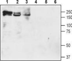
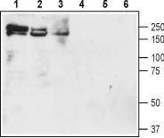
- Western blot analysis of rat brain membranes (lanes 1 and 4), mouse brain membranes (lanes 2 and 5) and rat lung lysate (lanes 3 and 6): 1-3. Anti-NKCC1 (extracellular) antibody, (1:200). 4-6. Anti-NKCC1 (extracellular) antibody, preincubated with the control peptide antigen.
- Validation comment
- WB
Supportive validation
- Submitted by
- OriGene (provider)
- Main image
- Experimental details
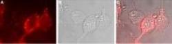
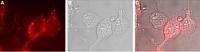
- Expression of NKCC1 in human U-87 MG cells. Immunocytochemical staining of live intact human U-87 MG glioblastoma cells. A. Extracellular staining of cells with Anti-NKCC1 (extracellular) antibody, (1:50), followed by goat anti-rabbit-AlexaFluor-594 secondary antibody (red). B. Live view of the cells. C. Merge of the two pictures.
- Validation comment
- IF
Supportive validation
- Submitted by
- OriGene (provider)
- Main image
- Experimental details
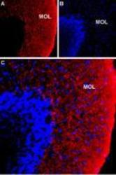
- Expression of NKCC1 in rat cerebellum. Immunohistochemical staining of immersion-fixed, free floating rat brain frozen sections using Anti-NKCC1 (extracellular) antibody, (1:100). A. NKCC1 staining (red) is detected in the cerebellar molecular layer (Mol). B. DAPI counterstain (blue) reveals the outline of cerebellar layers. C. Merge of the two images.
- Validation comment
- IHC
