Explore
 Validate
 Learn
Learn Western blot
Antibody data
- Antibody Data
- Antigen structure
- References [0]
- Comments [0]
- Validations
- Western blot [2]
- Immunocytochemistry [1]
- Immunohistochemistry [1]
- Flow cytometry [1]
Submit
Validation data
Reference
Comment
Report error
- Product number
- ANT-071-25UL - Provider product page

- Provider
- Invitrogen Antibodies
- Product name
- NKCC1 (SLC12A2) (extracellular) Polyclonal Antibody
- Antibody type
- Polyclonal
- Antigen
- Other
- Reactivity
- Human, Mouse, Rat
- Host
- Rabbit
- Isotype
- IgG
- Vial size
- 25 µL
- Concentration
- 0.6 mg/mL
- Storage
- -20° C, Avoid Freeze/Thaw Cycles
No comments: Submit comment
Supportive validation
- Submitted by
- Invitrogen Antibodies (provider)
- Main image

- Experimental details
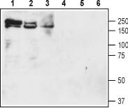
- Western blot analysis of rat brain membranes (lanes 1 and 4), mouse brain membranes (lanes 2 and 5) and rat lung lysate (lanes 3 and 6): - 1-3. Anti-NKCC1 (SLC12A2) (extracellular) Antibody (#ANT-071), (1:200).4-6. Anti-NKCC1 (SLC12A2) (extracellular) Antibody , preincubated with NKCC1/SLC12A2 (extracellular) Blocking Peptide (#BLP-NT071).

- Submitted by
- Invitrogen Antibodies (provider)
- Main image

- Experimental details
- Western blot analysis of rat brain membranes (lanes 1 and 4), mouse brain membranes (lanes 2 and 5) and rat lung lysate (lanes 3 and 6): - 1-3. Anti-NKCC1 (SLC12A2) (extracellular) Antibody (#ANT-071), (1:200).4-6. Anti-NKCC1 (SLC12A2) (extracellular) Antibody , preincubated with NKCC1/SLC12A2 (extracellular) Blocking Peptide (#BLP-NT071).
Supportive validation
- Submitted by
- Invitrogen Antibodies (provider)
- Main image
- Experimental details
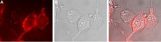
- Expression of NKCC1 in human U-87 MG cells - Cell surface detection of NKCC1 in live intact human U-87 MG glioblastoma cells. A. Extracellular staining of cells with Anti-NKCC1 (SLC12A2) (extracellular) Antibody (#ANT-071), (1:50), followed by goat Anti-rabbit-AlexaFluor-594 secondary Antibody (red). B. Live view of the cells. C. Merge of the two pictures.
Supportive validation
- Submitted by
- Invitrogen Antibodies (provider)
- Main image
- Experimental details
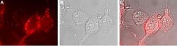
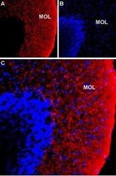
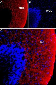
- Expression of NKCC1 in rat cerebellum - Immunohistochemical staining of immersion-fixed, free floating rat brain frozen sections using Anti-NKCC1 (SLC12A2) (extracellular) Antibody (#ANT-071), (1:100). A. NKCC1 staining (red) is detected in the cerebellar molecular layer (Mol). B. DAPI counterstain (blue) reveals the outline of cerebellar layers. C. Merge of the two images.
Supportive validation
- Submitted by
- Invitrogen Antibodies (provider)
- Main image
- Experimental details
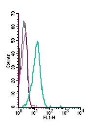
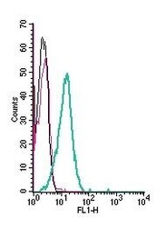
- Cell surface detection of NKCC1 by indirect flow cytometry in live intact human THP-1 monocytic leukemia cells: - (black line) cells. (red) Cells + goat- Anti-rabbit-FITC. (green) Cells + Anti-NKCC1 (SLC12A2) (extracellular) Antibody (#ANT-071), (2.5μg) + goat- Anti-rabbit-FITC.