Explore
 Validate
 Learn
Learn Western blot
Antibody data
- Antibody Data
- Antigen structure
- References [0]
- Comments [0]
- Validations
- Western blot [3]
- Immunohistochemistry [9]
Submit
Validation data
Reference
Comment
Report error
- Product number
- NBP1-81539 - Provider product page

- Provider
- Novus Biologicals
- Proper citation
- Novus Cat#NBP1-81539, RRID:AB_11017101
- Product name
- Rabbit Polyclonal EFR3A Antibody
- Antibody type
- Polyclonal
- Description
- Immunogen affinity purified. Specificity of human, mouse, rat EFR3A antibody verified on a Protein Array containing target protein plus 383 other non-specific proteins.
- Reactivity
- Human, Mouse, Rat
- Host
- Rabbit
- Isotype
- IgG
- Vial size
- 0.1 ml
- Storage
- Store at 4C short term. Aliquot and store at -20C long term. Avoid freeze-thaw cycles.
No comments: Submit comment
Supportive validation
- Submitted by
- Novus Biologicals (provider)
- Main image

- Experimental details
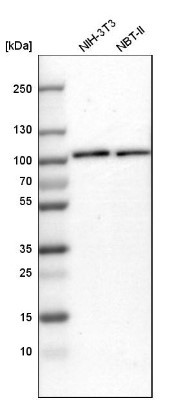
- Western Blot: EFR3A Antibody [NBP1-81539] - Analysis in mouse cell line NIH-3T3 and rat cell line NBT-II.
- Submitted by
- Novus Biologicals (provider)
- Main image
- Experimental details
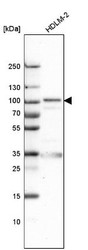
- Western Blot: EFR3A Antibody [NBP1-81539] - Analysis in human cell line HDLM-2.
- Submitted by
- Novus Biologicals (provider)
- Main image
- Experimental details
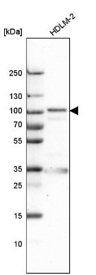
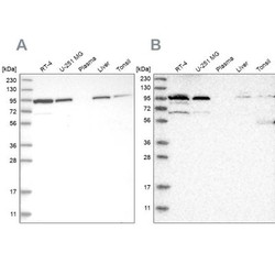
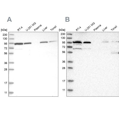
- Western Blot: EFR3A Antibody [NBP1-81539] - Analysis using Anti-EFR3A antibody NBP1-81539 (A) shows similar pattern to independent antibody NBP1-81538 (B).
Supportive validation
- Submitted by
- Novus Biologicals (provider)
- Main image
- Experimental details
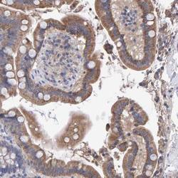
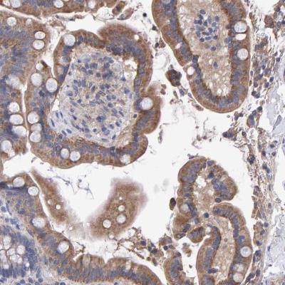
- Immunohistochemistry-Paraffin: EFR3A Antibody [NBP1-81539] - Staining of human colon shows moderate cytoplasmic positivity in glandular cells.
- Submitted by
- Novus Biologicals (provider)
- Main image
- Experimental details
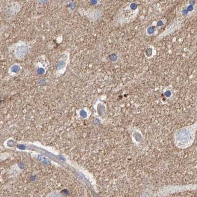
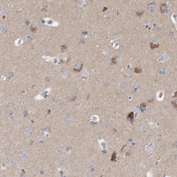
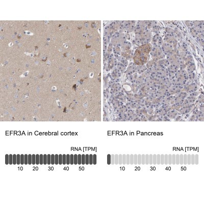
- Immunohistochemistry-Paraffin: EFR3A Antibody [NBP1-81539] - Staining of human cerebral cortex shows high expression.
- Submitted by
- Novus Biologicals (provider)
- Main image
- Experimental details
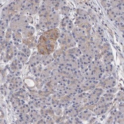
- Immunohistochemistry-Paraffin: EFR3A Antibody [NBP1-81539] - Staining of human pancreas shows low expression as expected.
- Submitted by
- Novus Biologicals (provider)
- Main image
- Experimental details
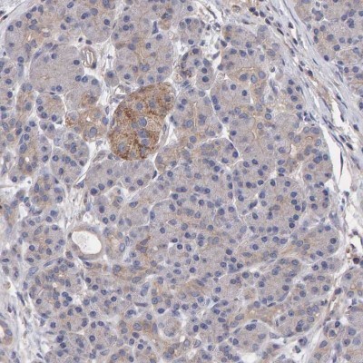
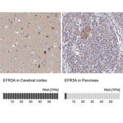
- Immunohistochemistry-Paraffin: EFR3A Antibody [NBP1-81539] - Staining in human cerebral cortex and pancreas tissues using anti-EFR3A antibody. Corresponding EFR3A RNA-seq data are presented for the same tissues.
- Submitted by
- Novus Biologicals (provider)
- Main image
- Experimental details
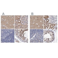
- Immunohistochemistry-Paraffin: EFR3A Antibody [NBP1-81539] - Staining of human cerebral cortex, kidney, lymph node and testis using Anti-EFR3A antibody NBP1-81539 (A) shows similar protein distribution across tissues to independent antibody NBP1-81540 (B).

- Submitted by
- Novus Biologicals (provider)
- Main image
- Experimental details
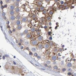
- Immunohistochemistry-Paraffin: EFR3A Antibody [NBP1-81539] - Staining of human testis.
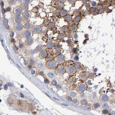
- Submitted by
- Novus Biologicals (provider)
- Main image
- Experimental details
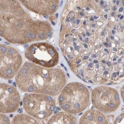
- Immunohistochemistry-Paraffin: EFR3A Antibody [NBP1-81539] - Staining of human kidney.

- Submitted by
- Novus Biologicals (provider)
- Main image
- Experimental details
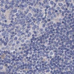
- Immunohistochemistry-Paraffin: EFR3A Antibody [NBP1-81539] - Staining of human lymph node.
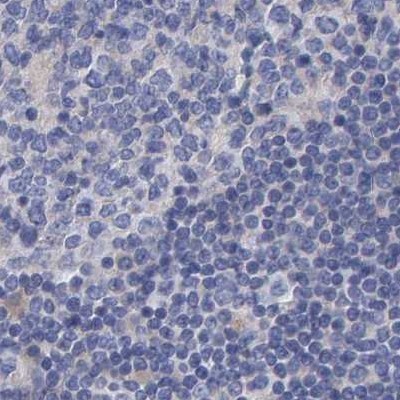
- Submitted by
- Novus Biologicals (provider)
- Main image

- Experimental details
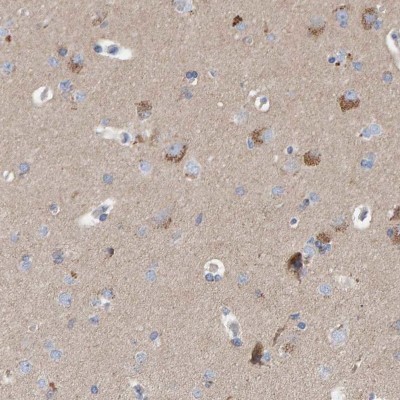
- Immunohistochemistry-Paraffin: EFR3A Antibody [NBP1-81539] - Staining of human cerebral cortex using Anti-EFR3A antibody NBP1-81539.