Explore
 Validate
 Learn
Learn Immunocytochemistry
 Immunohistochemistry
Antibody data
- Antibody Data
- Antigen structure
- References [3]
- Comments [0]
- Validations
- Immunocytochemistry [1]
- Other assay [1]
Submit
Validation data
Reference
Comment
Report error
- Product number
- PA1-930 - Provider product page

- Provider
- Invitrogen Antibodies
- Product name
- KCNQ3 Polyclonal Antibody
- Antibody type
- Polyclonal
- Antigen
- Other
- Description
- PA1-930 detects KCNQ3 from mouse, human and rat samples. This antibody is specific for KCNQ3 and does not detect KCNQ1, KCNQ2, KCNQ4 or KCNQ5. PA1-930 has been successfully used in immunofluorescence, immunohistochemistry and immunocytochemistry procedures. Immunohistochemical staining using PA1-930 yielded a strong signal mainly in interneurons and astrocytes in the dentate region of rat hippocampal samples. PA1-930 immunogen is a GST fusion protein encoding the first 71 amino acids of rat KCNQ3.
- Reactivity
- Human, Mouse, Rat
- Host
- Rabbit
- Isotype
- IgG
- Vial size
- 100 μg
- Concentration
- 1 mg/mL
- Storage
- -20°C, Avoid Freeze/Thaw Cycles
Submitted references A novel homozygous KCNQ3 loss-of-function variant causes non-syndromic intellectual disability and neonatal-onset pharmacodependent epilepsy.
Loss of the m-AAA protease subunit AFG₃L₂ causes mitochondrial transport defects and tau hyperphosphorylation.
Antibodies and a cysteine-modifying reagent show correspondence of M current in neurons to KCNQ2 and KCNQ3 K+ channels.
Lauritano A, Moutton S, Longobardi E, Tran Mau-Them F, Laudati G, Nappi P, Soldovieri MV, Ambrosino P, Cataldi M, Jouan T, Lehalle D, Maurey H, Philippe C, Miceli F, Vitobello A, Taglialatela M
Epilepsia open 2019 Sep;4(3):464-475
Epilepsia open 2019 Sep;4(3):464-475
Loss of the m-AAA protease subunit AFG₃L₂ causes mitochondrial transport defects and tau hyperphosphorylation.
Kondadi AK, Wang S, Montagner S, Kladt N, Korwitz A, Martinelli P, Herholz D, Baker MJ, Schauss AC, Langer T, Rugarli EI
The EMBO journal 2014 May 2;33(9):1011-26
The EMBO journal 2014 May 2;33(9):1011-26
Antibodies and a cysteine-modifying reagent show correspondence of M current in neurons to KCNQ2 and KCNQ3 K+ channels.
Roche JP, Westenbroek R, Sorom AJ, Hille B, Mackie K, Shapiro MS
British journal of pharmacology 2002 Dec;137(8):1173-86
British journal of pharmacology 2002 Dec;137(8):1173-86
No comments: Submit comment
Supportive validation
- Submitted by
- Invitrogen Antibodies (provider)
- Main image

- Experimental details
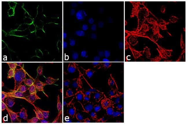
- Immunofluorescence analysis of KCNQ3 was performed using 70% confluent log phase RSC-96 cells. The cells were fixed with 4% paraformaldehyde for 10 minutes, permeabilized with 0.1% Triton X-100 for 10 minutes, and blocked with 1% BSA for 1 hour at room temperature. The cells were labeled with KCNQ3 Rabbit Polyclonal Antibody (Product # PA1-930) at 2 µg/mL in 0.1% BSA and incubated for 3 hours at room temperature and then labeled with Goat anti-Rabbit IgG (Heavy Chain) Superclonal Secondary Antibody, Alexa Fluor® 488 conjugate (Product # A27034) at a dilution of 1:2000 for 45 minutes at room temperature (Panel a: green). Nuclei (Panel b: blue) were stained with SlowFade® Gold Antifade Mountant with DAPI (Product # S36938). F-actin (Panel c: red) was stained with Alexa Fluor® 555 Rhodamine Phalloidin (Product # R415, 1:300). Panel d represents the merged image showing membranous localization. Panel e shows the no primary antibody control. The images were captured at 60X magnification.
Supportive validation
- Submitted by
- Invitrogen Antibodies (provider)
- Main image
- Experimental details
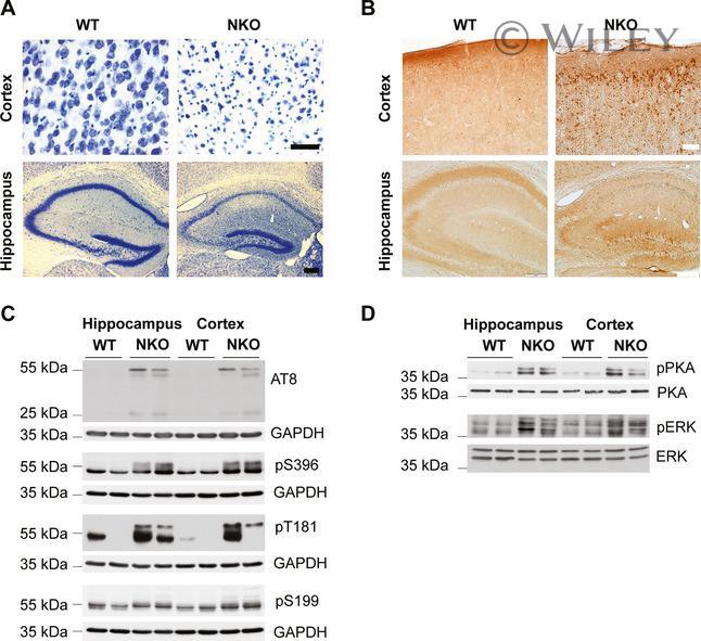
- Deletion of Afg3l2 in adult forebrain neurons causes tau hyperphosphorylation and activation of PKA and ERK 1/2 kinases Brain coronal sections of Afg3l2 forebrain neuronal specific knockout ( Afg3l2 fl/fl ; CamKIIalpha-Cre positive; NKO ) and littermate control ( Afg3l2 fl/fl ; CamKIIalpha-Cre negative, WT ) at 8 weeks of age were stained with Nissl solution. Pronounced neuronal degeneration is observed in the cerebral cortex and hippocampus. AT8 immunohistochemistry staining of adjacent sections from (A) shows accumulation of hyperphosphorylated tau in neuronal cell bodies in both cerebral cortex and hippocampus of Afg3l2 NKO . Western blot analysis of hippocampus and cerebral cortex lysates from Afg3l2 NKO and WT littermates using the indicated antibodies detects increased phosphorylation of tau. Western blot analysis of hippocampus and cerebral cortex lysates from Afg3l2 NKO and WT littermates using the indicated antibodies show activation of PKA and ERK1/2 kinases. Data information: Scale bar in (A) 50 mum (cortex) and 200 mum (hippocampus), in (B) 50 mum (cortex) and 200 mum (hippocampus). Source data are available online for this figure.