Explore
 Validate
 Learn
Learn Western blot
 Immunocytochemistry
Antibody data
- Antibody Data
- Antigen structure
- References [3]
- Comments [0]
- Validations
- Immunocytochemistry [4]
- Immunohistochemistry [1]
- Flow cytometry [1]
- Other assay [1]
Submit
Validation data
Reference
Comment
Report error
- Product number
- PA5-23034 - Provider product page

- Provider
- Invitrogen Antibodies
- Product name
- PKM2 Polyclonal Antibody
- Antibody type
- Polyclonal
- Antigen
- Synthetic peptide
- Description
- The target sequence has 95% sequence homology with bovine.
- Reactivity
- Human, Mouse, Rat, Bovine
- Host
- Rabbit
- Isotype
- IgG
- Vial size
- 100 μL
- Concentration
- 1 mg/mL
- Storage
- -20°C, Avoid Freeze/Thaw Cycles
Submitted references Longitudinal in vivo bioimaging of hepatocyte transcription factor activity following cholestatic liver injury in mice.
Breast cancer stem cells rely on fermentative glycolysis and are sensitive to 2-deoxyglucose treatment.
p63 isoforms regulate metabolism of cancer stem cells.
Delhove JM, Buckley SM, Perocheau DP, Karda R, Arbuthnot P, Henderson NC, Waddington SN, McKay TR
Scientific reports 2017 Feb 3;7:41874
Scientific reports 2017 Feb 3;7:41874
Breast cancer stem cells rely on fermentative glycolysis and are sensitive to 2-deoxyglucose treatment.
Ciavardelli D, Rossi C, Barcaroli D, Volpe S, Consalvo A, Zucchelli M, De Cola A, Scavo E, Carollo R, D'Agostino D, Forlì F, D'Aguanno S, Todaro M, Stassi G, Di Ilio C, De Laurenzi V, Urbani A
Cell death & disease 2014 Jul 17;5(7):e1336
Cell death & disease 2014 Jul 17;5(7):e1336
p63 isoforms regulate metabolism of cancer stem cells.
D'Aguanno S, Barcaroli D, Rossi C, Zucchelli M, Ciavardelli D, Cortese C, De Cola A, Volpe S, D'Agostino D, Todaro M, Stassi G, Di Ilio C, Urbani A, De Laurenzi V
Journal of proteome research 2014 Apr 4;13(4):2120-36
Journal of proteome research 2014 Apr 4;13(4):2120-36
No comments: Submit comment
Supportive validation
- Submitted by
- Invitrogen Antibodies (provider)
- Main image

- Experimental details
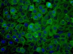
- Immunocytochemistry analysis of PKM2 in HeLa cells. Samples were incubated in PKM2 polyclonal antibody (Product # PA5-23034).

- Submitted by
- Invitrogen Antibodies (provider)
- Main image
- Experimental details
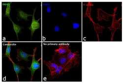
- Immunofluorescence analysis of PKM2 was performed using 70% confluent log phase MDA-MB-231 cells. The cells were fixed with 4% paraformaldehyde for 10 minutes, permeabilized with 0.1% Triton™ X-100 for 15 minutes, and blocked with 1% BSA for 1 hour at room temperature. The cells were labeled with PKM2 Polyclonal Antibody (Product # PA5-23034) at 1:100 dilution in 0.1% BSA, incubated at 4 degree Celsius overnight and then labeled with Goat anti-Rabbit IgG (H+L) Superclonal™ Secondary Antibody, Alexa Fluor® 488 conjugate (Product # A27034) at a dilution of 1:2000 for 45 minutes at room temperature (Panel a: green). Nuclei (Panel b: blue) were stained with SlowFade® Gold Antifade Mountant with DAPI (Product # S36938). F-actin (Panel c: red) was stained with Rhodamine Phalloidin (Product # R415, 1:300). Panel d represents the merged image showing cytoplasmic and nuclear localization. Panel e represents control cells with no primary antibody to assess background. The images were captured at 60X magnification.

- Submitted by
- Invitrogen Antibodies (provider)
- Main image

- Experimental details
- Immunofluorescence analysis of PKM2 was performed using 70% confluent log phase MDA-MB-231 cells. The cells were fixed with 4% paraformaldehyde for 10 minutes, permeabilized with 0.1% Triton™ X-100 for 15 minutes, and blocked with 1% BSA for 1 hour at room temperature. The cells were labeled with PKM2 Polyclonal Antibody (Product # PA5-23034) at 1:100 dilution in 0.1% BSA, incubated at 4 degree Celsius overnight and then labeled with Goat anti-Rabbit IgG (Heavy Chain) Superclonal™ Secondary Antibody, Alexa Fluor® 488 conjugate (Product # A27034) at a dilution of 1:2000 for 45 minutes at room temperature (Panel a: green). Nuclei (Panel b: blue) were stained with SlowFade® Gold Antifade Mountant with DAPI (Product # S36938). F-actin (Panel c: red) was stained with Rhodamine Phalloidin (Product # R415, 1:300). Panel d represents the merged image showing cytoplasmic and nuclear localization. Panel e represents control cells with no primary antibody to assess background. The images were captured at 60X magnification.

- Submitted by
- Invitrogen Antibodies (provider)
- Main image
- Experimental details
- Immunocytochemistry analysis of PKM2 in HeLa cells. Samples were incubated in PKM2 polyclonal antibody (Product # PA5-23034).

Supportive validation
- Submitted by
- Invitrogen Antibodies (provider)
- Main image

- Experimental details
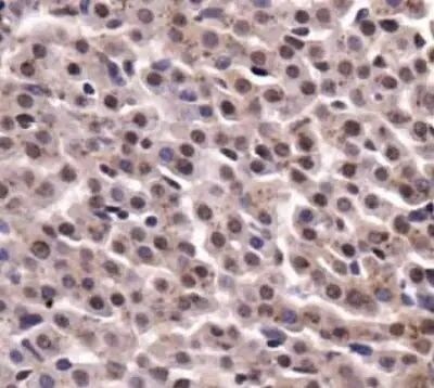
- Immunohistochemical analysis of PKM2 in mouse liver tissue. Samples were incubated in PKM2 polyclonal antibody (Product # PA5-23034).
Supportive validation
- Submitted by
- Invitrogen Antibodies (provider)
- Main image
- Experimental details
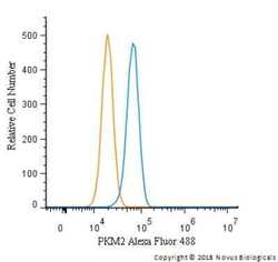
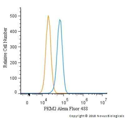
- Flow cytometry of PKM2 in HeLa cells (blue) and a matched isotype control (orange). Samples were incubated in PKM2 polyclonal antibody (Product # PA5-23034) using a dilution of 5 µg/mL for 30 minutes at room temperature. Cells were fixed with 4% PFA and then permeabilized with 0.1% saponin. Both antibodies were conjugated to Alexa Fluor 488.
Supportive validation
- Submitted by
- Invitrogen Antibodies (provider)
- Main image
- Experimental details
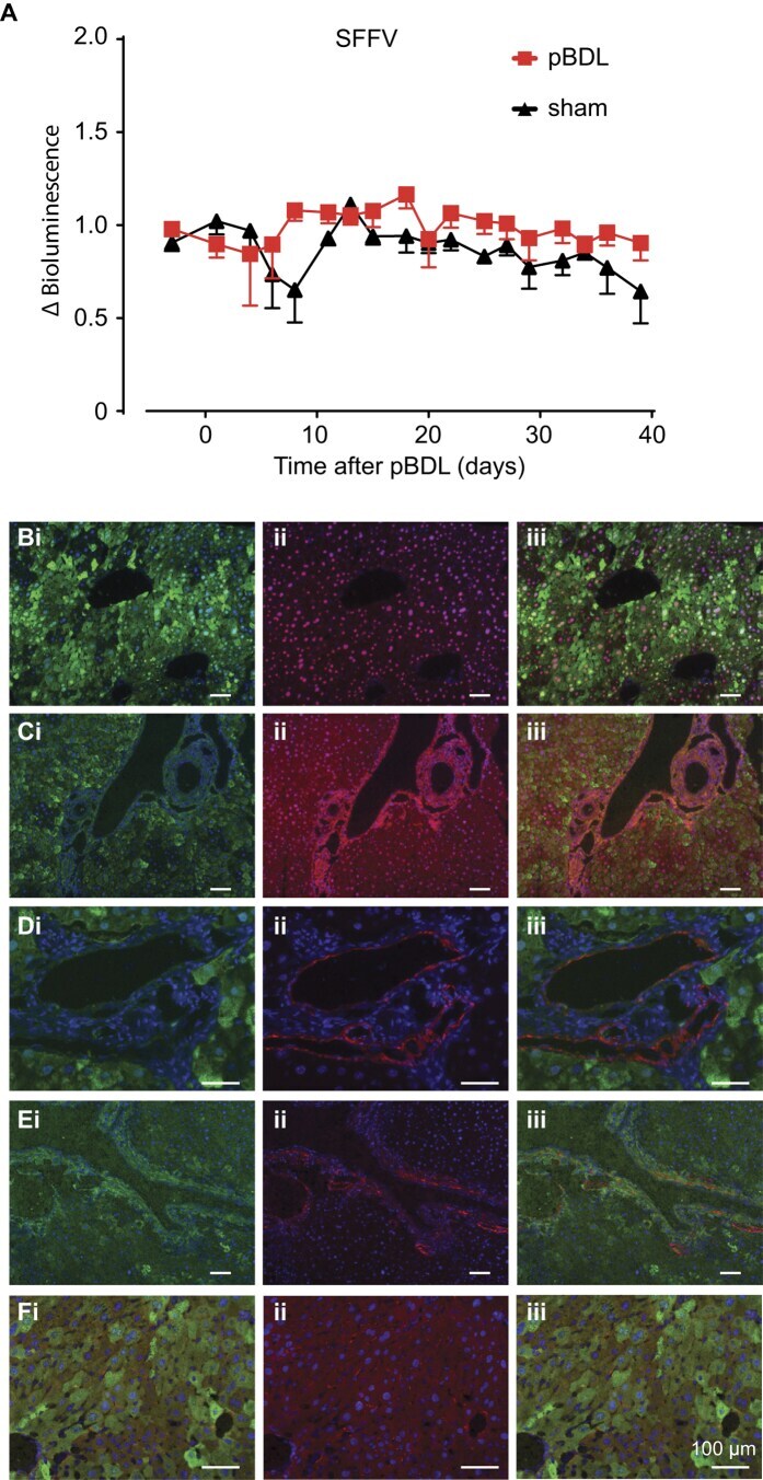
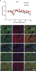
- Figure 3 Reporter gene expression is stable and restricted to hepatocytes after pBDL following neonatal intravascular administration of lentiviral vectors. SFFV-FLuc/eGFP lentivirus was administered to P1 neonatal mice by intravascular (i.v.) injection and then subjected to pBDL or sham pBDL in adulthood. ( A ) Mice were subjected to continued luciferase bioimaging over 40 days where no change in luciferase activity was observed over time or between pBDL and sham groups (n = 10 pBDL, 5 sham, not significant, Student's t-test). Mice were sacrificed 90 days after pBDL and co-immunostained for GFP and markers of ( Bi-iii ) hepatocytes; HNF4alpha, ( C-iii ) biliary epithelia; CK7, ( Di-iii ) hepatic progenitors; PKM2, ( Ei-iii ) myofibroblasts; alphaSMA and ( Fi-iii ) hepatic stellate cells; GFAP (all groups n = 3-6).