Explore
 Validate
 Learn
Learn Western blot
Antibody data
- Antibody Data
- Antigen structure
- References [1]
- Comments [0]
- Validations
- Western blot [6]
- Immunocytochemistry [1]
- Immunohistochemistry [1]
- Other assay [1]
Submit
Validation data
Reference
Comment
Report error
- Product number
- PA5-28623 - Provider product page

- Provider
- Invitrogen Antibodies
- Product name
- PKM2 Polyclonal Antibody
- Antibody type
- Polyclonal
- Antigen
- Recombinant protein fragment
- Description
- Recommended positive controls: 293T, A431, H1299, HeLaS3, HepG2, Molt-4, Raji, mouse brain, rat brain. Predicted reactivity: Human (99%), Mouse (97%), Rat (97%), Xenopus laevis (88%), Rabbit (96%), Chicken (87%), Bovine (96%). Store product as a concentrated solution. Centrifuge briefly prior to opening the vial.
- Reactivity
- Human, Mouse, Rat
- Host
- Rabbit
- Isotype
- IgG
- Vial size
- 100 µL
- Concentration
- 0.14 mg/mL
- Storage
- Store at 4°C short term. For long term storage, store at -20°C, avoiding freeze/thaw cycles.
Submitted references SEMG1/2 augment energy metabolism of tumor cells.
Shuvalov O, Kizenko A, Petukhov A, Fedorova O, Daks A, Bottrill A, Snezhkina AV, Kudryavtseva AV, Barlev N
Cell death & disease 2020 Dec 11;11(12):1047
Cell death & disease 2020 Dec 11;11(12):1047
No comments: Submit comment
Supportive validation
- Submitted by
- Invitrogen Antibodies (provider)
- Main image
- Experimental details
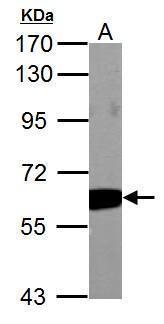
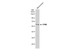
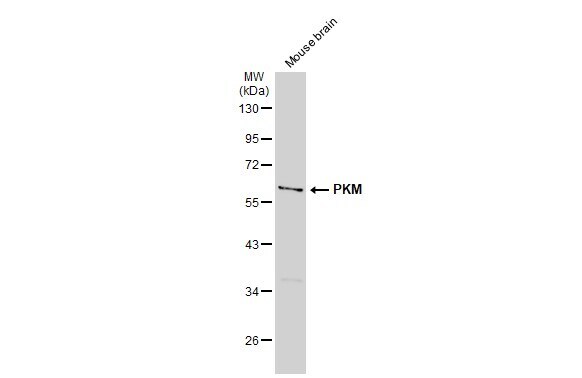
- Western blot analysis of Pyruvate Kinase (muscle) using 50 µg of mouse brain lysate. Samples were loaded onto a 7.5% SDS-PAGE gel and probed with a Pyruvate Kinase (muscle) polyclonal antibody (Product # PA5-28623) at a dilution of 1:1000.

- Submitted by
- Invitrogen Antibodies (provider)
- Main image
- Experimental details
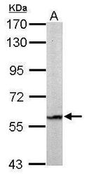
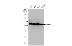
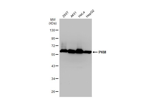
- Western blot analysis of Pyruvate Kinase (muscle) using 30µg of A) 293T (B) A431 (C) H1299 (D) HeLa S3 (E) HepG2 (F) MOLT4 and G) Raji lysate. Samples were loaded onto a 7.5% SDS-PAGE gel and probed with a Pyruvate Kinase (muscle) polyclonal antibody (Product # PA5-28623) at a dilution of 1:1000.
- Submitted by
- Invitrogen Antibodies (provider)
- Main image
- Experimental details
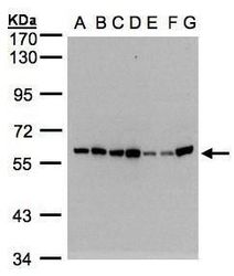
- Western Blot using PKM2 Polyclonal Antibody (Product # PA5-28623). Various whole cell extracts (30 µg) were separated by 10% SDS-PAGE, and the membrane was blotted with PKM2 Polyclonal Antibody (Product # PA5-28623) diluted at 1:1,000. The HRP-conjugated anti-rabbit IgG antibody was used to detect the primary antibody.
- Submitted by
- Invitrogen Antibodies (provider)
- Main image
- Experimental details
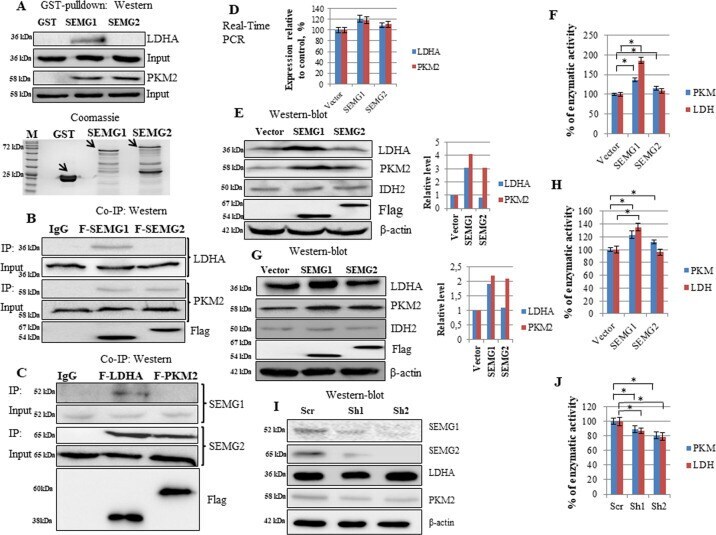
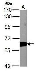
- Pyruvate Kinase (muscle) antibody detects PKM2 protein by Western blot analysis. A. 50 µg Rat brain lysate/extract.7.5 % SDS-PAGE. Pyruvate Kinase (muscle) antibody PKM2 Polyclonal Antibody (Product # PA5-28623) dilution: 1:1,000.
- Submitted by
- Invitrogen Antibodies (provider)
- Main image
- Experimental details
- Western blot analysis of PKM2 was performed by separating 50 µg of mouse tissue extract by 10% SDS-PAGE. Proteins were transferred to a membrane and probed with a PKM2 Polyclonal Antibody (Product # PA5-28623) at a dilution of 1:1000. The HRP-conjugated anti-rabbit IgG antibody was used to detect the primary antibody.
- Submitted by
- Invitrogen Antibodies (provider)
- Main image
- Experimental details
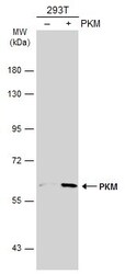
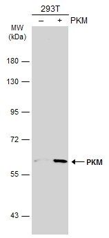
- Western Blot analysis of PKM2 was performed by separating 30 µg of non-transfected (–) and transfected (+) 293T whole cell extracts by 5% SDS-PAGE. Proteins were transferred to a membrane and probed with a PKM2 Polyclonal Antibody (Product # PA5-28623) at a dilution of 1:500. The HRP-conjugated anti-rabbit IgG antibody was used to detect the primary antibody.
Supportive validation
- Submitted by
- Invitrogen Antibodies (provider)
- Main image

- Experimental details
- PKM2 Polyclonal Antibody detects PKM protein at cytoplasm by immunofluorescent analysis. Sample: HeLa cells were fixed in 4% paraformaldehyde at RT for 15 min. Green: PKM stained by PKM2 Polyclonal Antibody (Product # PA5-28623) diluted at 1:500.

Supportive validation
- Submitted by
- Invitrogen Antibodies (provider)
- Main image

- Experimental details
- PKM2 Polyclonal Antibody detects PKM protein at cytoplasm by immunohistochemical analysis. Sample: Paraffin-embedded human lung cancer. PKM stained by PKM2 Polyclonal Antibody (Product # PA5-28623) diluted at 1:500. Antigen Retrieval: Citrate buffer, pH 6.0, 15 min.

Supportive validation
- Submitted by
- Invitrogen Antibodies (provider)
- Main image
- Experimental details
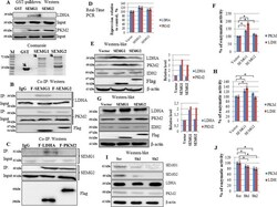
- Fig. 4 SEMG1 and SEMG2 interact with PKM2 and LDHA, upregulates their protein level and enzymatic activity. Recombinant SEMG1 and SEMG2 bind PKM2, whereas SEMG1 only binds LDHA in GST pull-down assay followed by western-blotting. B 3xFlag-tagged SEMG1 and SEMG2 bind PKM2, whereas SEMG1 only binds LDHA in co-immunoprecipitation. C 3xFlag-tagged PKM2 interacts with both endogenous SEMG1 and SEMG2 in H520 cells, whereas 3xFlag-tagged LDHA binds SEMG1 only (co-immunoprecipitation). D Overexpression of SEMG1 and SEMG2 in H1299 cell line does not alters the mRNA levels of PKM2 and LDHA (Real-Time PCR). The stable overexpression of SEMG1 and SEMG2 increase the protein level and enzymatic activity of PKM2, whereas the overexpression of SEMG1 only elevates the protein level and enzymatic activity of LDHA in E , F H1299 cells and G , H Mia-Paca 2 cells. Knockdown of SEMG1 and SEMG2 in H520 cells decreases the protein level ( I ) and enzymatic activity ( J ) of PKM2 and LDHA. Three biological replicates were used for all quantifications, data are presented as mean +- S.D., * P < 0.05.