Explore
 Validate
 Learn
Learn Western blot
Antibody data
- Antibody Data
- Antigen structure
- References [1]
- Comments [0]
- Validations
- Western blot [3]
- Immunocytochemistry [2]
- Immunohistochemistry [2]
Submit
Validation data
Reference
Comment
Report error
- Product number
- AF7244 - Provider product page

- Provider
- R&D Systems
- Product name
- Human/Mouse/Rat PKM2 Antibody
- Antibody type
- Polyclonal
- Description
- Antigen Affinity-purified. Detects human, mouse, and rat PKM2 in Western blots and detects recombinant human PKM2 in direct ELISAs.
- Reactivity
- Human, Mouse, Rat
- Host
- Sheep
- Conjugate
- Unconjugated
- Antigen sequence
P14618- Isotype
- IgG
- Vial size
- 100 ug
- Concentration
- LYOPH
- Storage
- Use a manual defrost freezer and avoid repeated freeze-thaw cycles. 12 months from date of receipt, -20 to -70 °C as supplied. 1 month, 2 to 8 °C under sterile conditions after reconstitution. 6 months, -20 to -70 °C under sterile conditions after reconstitution.
Submitted references The airway epithelium undergoes metabolic reprogramming in individuals at high risk for lung cancer.
Rahman SMJ, Ji X, Zimmerman LJ, Li M, Harris BK, Hoeksema MD, Trenary IA, Zou Y, Qian J, Slebos RJ, Beane J, Spira A, Shyr Y, Eisenberg R, Liebler DC, Young JD, Massion PP
JCI insight 2016 Nov 17;1(19):e88814
JCI insight 2016 Nov 17;1(19):e88814
No comments: Submit comment
Supportive validation
- Submitted by
- R&D Systems (provider)
- Main image

- Experimental details
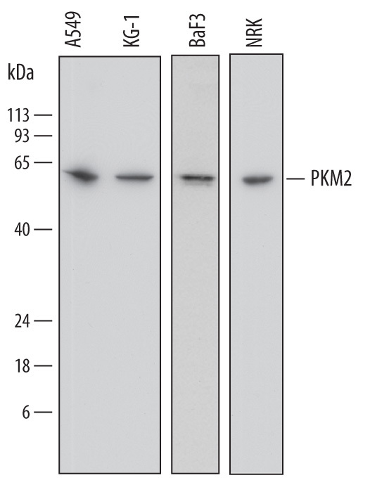
- Detection of Human, Mouse, and Rat PKM2 by Western Blot. Western blot shows lysates of A549 human lung carcinoma cell line, KG-1 human acute myelogenous leukemia cell line, BaF3 mouse pro-B cell line, and NRK rat normal kidney cell line. PVDF membrane was probed with 0.2 µg/mL of Sheep Anti-Human/Mouse/Rat PKM2 Antigen Affinity-purified Polyclonal Antibody (Catalog # AF7244) followed by HRP-conjugated Anti-Sheep IgG Secondary Antibody (Catalog # HAF016). A specific band was detected for PKM2 at approximately 60 kDa (as indicated). This experiment was conducted under reducing conditions and using Immunoblot Buffer Group 1.
- Submitted by
- R&D Systems (provider)
- Main image
- Experimental details
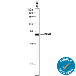
- Detection of Human PKM2 by Simple WesternTM. Simple Western lane view shows lysates of A549 human lung carcinoma cell line, loaded at 0.2 mg/mL. A specific band was detected for PKM2 at approximately 60 kDa (as indicated) using 2 µg/mL of Sheep Anti-Human/Mouse/Rat PKM2 Antigen Affinity-purified Polyclonal Antibody (Catalog # AF7244) followed by 1:50 dilution of HRP-conjugated Anti-Sheep IgG Secondary Antibody (Catalog # HAF016). This experiment was conducted under reducing conditions and using the 12-230 kDa separation system.
- Submitted by
- R&D Systems (provider)
- Main image
- Experimental details
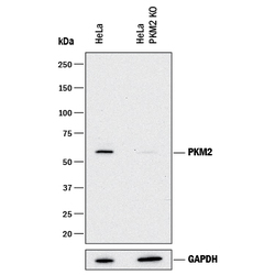
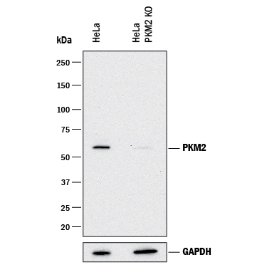
- Western Blot Shows Human PKM2 Specificity by Using Knockout Cell Line. Western blot shows lysates of HeLa human cervical epithelial carcinoma parental cell line and PKM2 knockout HeLa cell line (KO). PVDF membrane was probed with 1 µg/mL of Sheep Anti-Human/Mouse/Rat PKM2 Antigen Affinity-purified Polyclonal Antibody (Catalog # AF7244) followed by HRP-conjugated Anti-Sheep IgG Secondary Antibody (Catalog # HAF016). A specific band was detected for PKM2 at approximately 60 kDa (as indicated) in the parental HeLa cell line, but is not detectable in knockout HeLa cell line. GAPDH (Catalog # AF5718) is shown as a loading control. This experiment was conducted under reducing conditions and using Immunoblot Buffer Group 1.
Supportive validation
- Submitted by
- R&D Systems (provider)
- Main image

- Experimental details
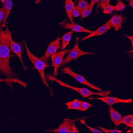
- PKM2 in RAW 264.7 Mouse Cell Line. PKM2 was detected in immersion fixed RAW 264.7 mouse monocyte/macrophage cell line using Sheep Anti-Human/Mouse/Rat PKM2 Antigen Affinity-purified Polyclonal Antibody (Catalog # AF7244) at 10 µg/mL for 3 hours at room temperature. Cells were stained using the NorthernLights™ 557-conjugated Anti-Sheep IgG Secondary Antibody (red; Catalog # NL010) and counterstained with DAPI (blue). Specific staining was localized to cytoplasm. View our protocol for Fluorescent ICC Staining of Cells on Coverslips.
- Submitted by
- R&D Systems (provider)
- Main image

- Experimental details
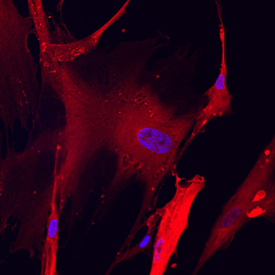
- PKM2 in Wi-38 Human Cell Line. PKM2 was detected in immersion fixed Wi-38 human lung fibroblast cell line using Sheep Anti-Human/Mouse/Rat PKM2 Antigen Affinity-purified Polyclonal Antibody (Catalog # AF7244) at 10 µg/mL for 3 hours at room temperature. Cells were stained using the NorthernLights™ 557-conjugated Anti-Sheep IgG Secondary Antibody (red; Catalog # NL010) and counterstained with DAPI (blue). Specific staining was localized to cytoplasm. View our protocol for Fluorescent ICC Staining of Cells on Coverslips.
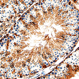
Supportive validation
- Submitted by
- R&D Systems (provider)
- Main image

- Experimental details
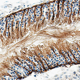
- PKM2 in Mouse Testis. PKM2 was detected in perfusion fixed frozen sections of mouse testis using Sheep Anti-Human/Mouse/Rat PKM2 Antigen Affinity-purified Polyclonal Antibody (Catalog # AF7244) at 1.7 µg/mL overnight at 4 °C. Tissue was stained using the Anti-Sheep HRP-DAB Cell & Tissue Staining Kit (brown; Catalog # CTS019) and counterstained with hematoxylin (blue). Specific staining was localized to Sertoli cells in testis. View our protocol for Chromogenic IHC Staining of Frozen Tissue Sections.
- Submitted by
- R&D Systems (provider)
- Main image

- Experimental details
- PKM2 in Rat Testis. PKM2 was detected in perfusion fixed frozen sections of rat testis using Sheep Anti-Human/Mouse/Rat PKM2 Antigen Affinity-purified Polyclonal Antibody (Catalog # AF7244) at 1.7 µg/mL overnight at 4 °C. Tissue was stained using the Anti-Sheep HRP-DAB Cell & Tissue Staining Kit (brown; Catalog # CTS019) and counterstained with hematoxylin (blue). Specific staining was localized to Sertoli cells in testis. View our protocol for Chromogenic IHC Staining of Frozen Tissue Sections.