Explore
 Validate
 Learn
LearnPA5-99434
antibody from Invitrogen Antibodies
Targeting: KEAP1
INrf2, KIAA0132, KLHL19, MGC10630, MGC1114, MGC20887, MGC4407, MGC9454
 Western blot
Antibody data
- Antibody Data
- Antigen structure
- References [1]
- Comments [0]
- Validations
- Western blot [3]
- Immunocytochemistry [3]
- Immunohistochemistry [14]
- Other assay [3]
Submit
Validation data
Reference
Comment
Report error
- Product number
- PA5-99434 - Provider product page

- Provider
- Invitrogen Antibodies
- Product name
- KEAP1 Polyclonal Antibody
- Antibody type
- Polyclonal
- Antigen
- Synthetic peptide
- Description
- Antibody detects endogenous levels of total Keap1.
- Reactivity
- Human, Mouse, Rat
- Host
- Rabbit
- Isotype
- IgG
- Vial size
- 100 μL
- Concentration
- 1 mg/mL
- Storage
- -20°C
Submitted references E3 ligase TRIM15 facilitates non-small cell lung cancer progression through mediating Keap1-Nrf2 signaling pathway.
Liang M, Wang L, Sun Z, Chen X, Wang H, Qin L, Zhao W, Geng B
Cell communication and signaling : CCS 2022 May 9;20(1):62
Cell communication and signaling : CCS 2022 May 9;20(1):62
No comments: Submit comment
Supportive validation
- Submitted by
- Invitrogen Antibodies (provider)
- Main image
- Experimental details
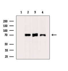
- Western blot analysis of KEAP1 in extracts from various samples. Samples were incubated with KEAP1 polyclonal antibody (Product # PA5-99434). Lane 1: Mouse brain, blocked with antigen-specific peptides; Lane 2: Mouse brain; Lane 3: Hepg2 cells (heat-shock treatment); Lane 4: Hela cells (heat-shock treatment).
- Submitted by
- Invitrogen Antibodies (provider)
- Main image
- Experimental details
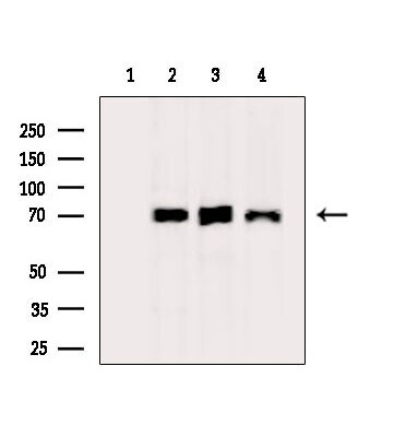
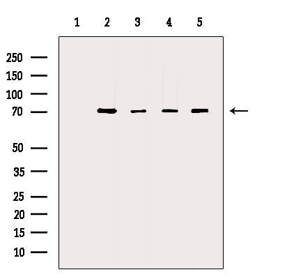
- Western blot analysis of KEAP1 in various samples (Lane 1: VERO treated with blocking peptide Lane 2: VERO Lane 3: mouse spleen Lane 4: 3T3 Lane 5: mouse muscle). Samples were incubated with KEAP1 polyclonal antibody (Product # PA5-99434).
- Submitted by
- Invitrogen Antibodies (provider)
- Main image

- Experimental details
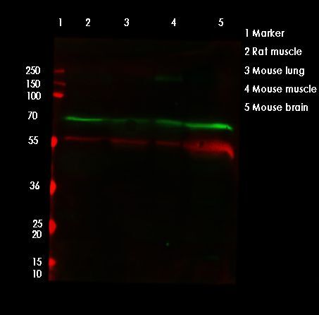
- Western blot analysis of KEAP1 in various lysate (Lane 1-2: merged signal, red and green). Samples were incubated with KEAP1 polyclonal antibody (Product # PA5-99434).
Supportive validation
- Submitted by
- Invitrogen Antibodies (provider)
- Main image
- Experimental details
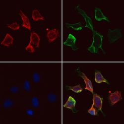
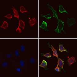
- Immunofluorescent analysis of KEAP1 in HepG2 cells. Samples were fixed with paraformaldehyde, permeabilized with 0.1% Triton X-100, blocked with 10% serum (45 min at 25°C), incubated with mouse anti-beta tubulin and KEAP1 polyclonal antibody (Product # PA5-99434) using a dilution of 1:200 (1 hr, 37°C), and followed by goat anti-rabbit IgG Alexa Fluor 594 (red) and goat anti-mouse IgG Alexa Fluor 488 (green).
- Submitted by
- Invitrogen Antibodies (provider)
- Main image
- Experimental details
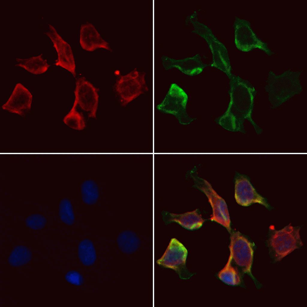
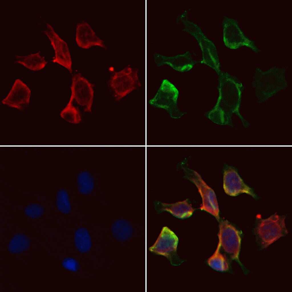
- Immunofluorescent analysis of KEAP1 in HepG2 cells. Samples were fixed with paraformaldehyde, permeabilized with 0.1% Triton X-100, blocked with 10% serum (45 min at 25°C), incubated with mouse anti-beta tubulin and KEAP1 polyclonal antibody (Product # PA5-99434) using a dilution of 1:200 (1 hr, 37°C), and followed by goat anti-rabbit IgG Alexa Fluor 594 (red) and goat anti-mouse IgG Alexa Fluor 488 (green).
- Submitted by
- Invitrogen Antibodies (provider)
- Main image
- Experimental details
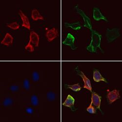
- Immunofluorescent analysis of KEAP1 in HepG2 cells. Samples were fixed with paraformaldehyde, permeabilized with 0.1% Triton X-100, blocked with 10% serum (45 min at 25°C), incubated with mouse anti-beta tubulin and KEAP1 polyclonal antibody (Product # PA5-99434) using a dilution of 1:200 (1 hr, 37°C), and followed by goat anti-rabbit IgG Alexa Fluor 594 (red) and goat anti-mouse IgG Alexa Fluor 488 (green).

Supportive validation
- Submitted by
- Invitrogen Antibodies (provider)
- Main image

- Experimental details
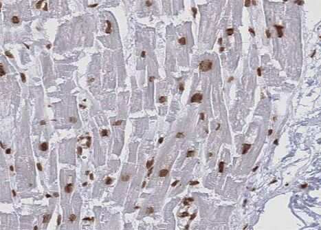
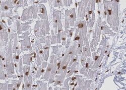
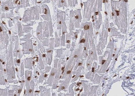
- Immunohistochemistry analysis of paraffin-embedded KEAP1 in human Heart muscle tissue sections. Antigen retrieval was performed using citrate buffer. Samples were blocked with blocking buffer (1.5 hr, 22°C), incubated with KEAP1 polyclonal antibody (Product # PA5-99434) using a dilution of 1:100 (1.5 hr, 22°C), followed by HRP conjugated goat anti-rabbit.
- Submitted by
- Invitrogen Antibodies (provider)
- Main image
- Experimental details
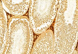
- Immunohistochemistry analysis of KEAP1 in rat testis tissue. The sample was formaldehyde fixed and a heat mediated antigen retrieval step in citrate buffer was performed. Samples were incubated with KEAP1 polyclonal antibody (Product # PA5-99434) using a dilution of 1:100 (4°C overnight) followed by HRP conjugated anti-Rabbit secondary antibody.
- Submitted by
- Invitrogen Antibodies (provider)
- Main image

- Experimental details
- Immunohistochemistry analysis of KEAP1 in mouse spleen tissue. The sample was formaldehyde fixed and a heat mediated antigen retrieval step in citrate buffer was performed. Samples were incubated with KEAP1 polyclonal antibody (Product # PA5-99434) using a dilution of 1:100 (4°C overnight) followed by HRP conjugated anti-Rabbit secondary antibody.

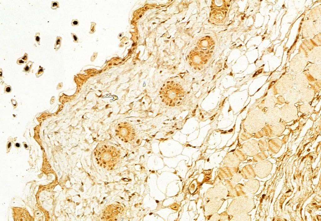
- Submitted by
- Invitrogen Antibodies (provider)
- Main image

- Experimental details
- Immunohistochemistry analysis of KEAP1 in mouse skin tissue. The sample was formaldehyde fixed and a heat mediated antigen retrieval step in citrate buffer was performed. Samples were incubated with KEAP1 polyclonal antibody (Product # PA5-99434) using a dilution of 1:100 (4°C overnight) followed by HRP conjugated anti-Rabbit secondary antibody.

- Submitted by
- Invitrogen Antibodies (provider)
- Main image
- Experimental details
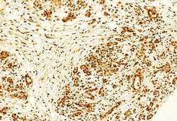
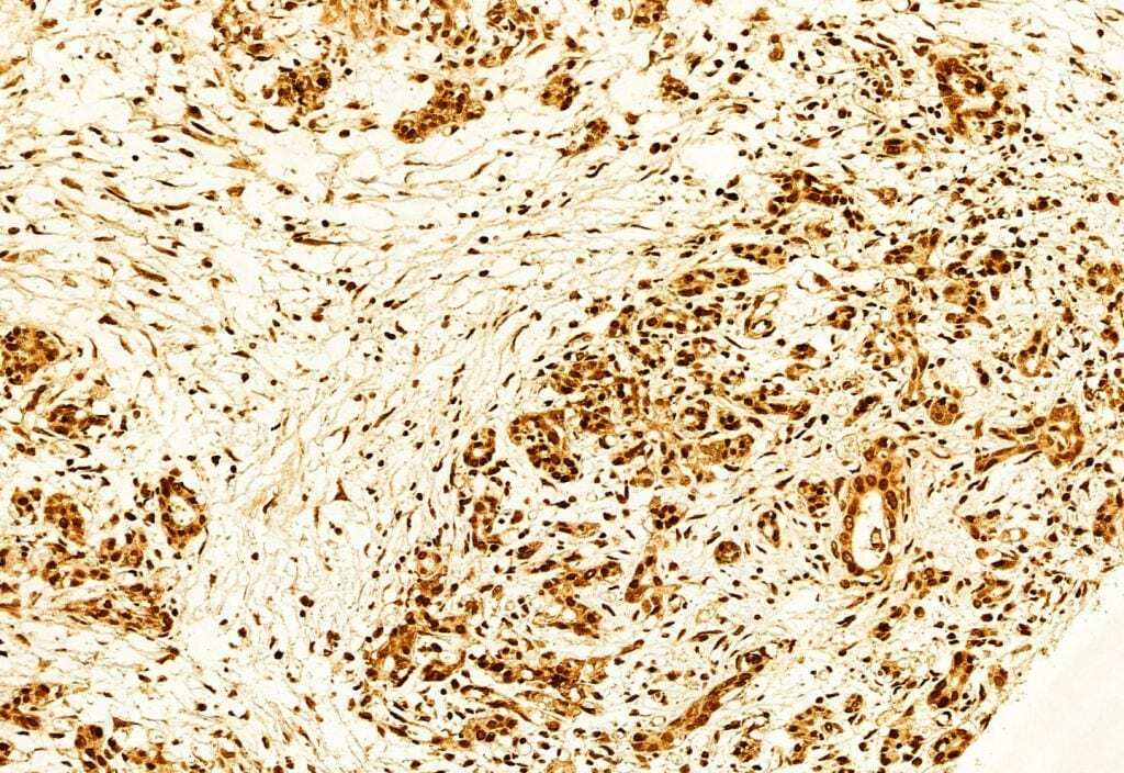
- Immunohistochemistry analysis of KEAP1 in human colorectal cancer. The sample was formaldehyde fixed and a heat mediated antigen retrieval step in citrate buffer was performed. Samples were incubated with KEAP1 polyclonal antibody (Product # PA5-99434) using a dilution of 1:100 (4°C overnight) followed by HRP conjugated anti-Rabbit secondary antibody.
- Submitted by
- Invitrogen Antibodies (provider)
- Main image
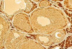
- Experimental details
- Immunohistochemistry analysis of KEAP1 in rat ovary tissue. The sample was formaldehyde fixed and a heat mediated antigen retrieval step in citrate buffer was performed. Samples were incubated with KEAP1 polyclonal antibody (Product # PA5-99434) using a dilution of 1:100 (4°C overnight) followed by HRP conjugated anti-Rabbit secondary antibody.
- Submitted by
- Invitrogen Antibodies (provider)
- Main image

- Experimental details
- Immunohistochemistry analysis of KEAP1 in rat skin tissue. The sample was formaldehyde fixed and a heat mediated antigen retrieval step in citrate buffer was performed. Samples were incubated with KEAP1 polyclonal antibody (Product # PA5-99434) using a dilution of 1:100 (4°C overnight) followed by HRP conjugated anti-Rabbit secondary antibody.

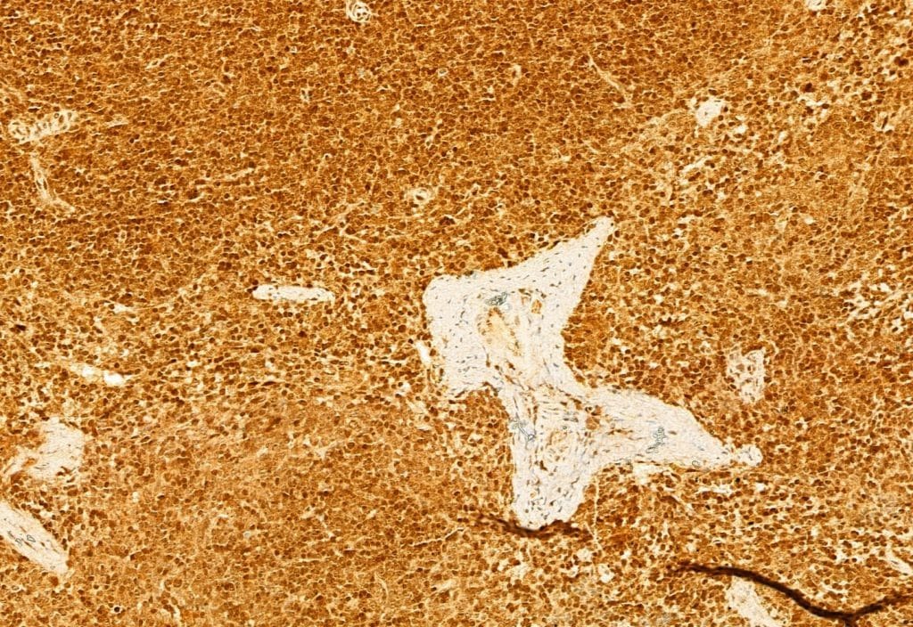
- Submitted by
- Invitrogen Antibodies (provider)
- Main image

- Experimental details
- Immunohistochemistry analysis of KEAP1 in mouse liver tissue. The sample was formaldehyde fixed and a heat mediated antigen retrieval step in citrate buffer was performed. Samples were incubated with KEAP1 polyclonal antibody (Product # PA5-99434) using a dilution of 1:100 (4°C overnight) followed by HRP conjugated anti-Rabbit secondary antibody.

- Submitted by
- Invitrogen Antibodies (provider)
- Main image

- Experimental details
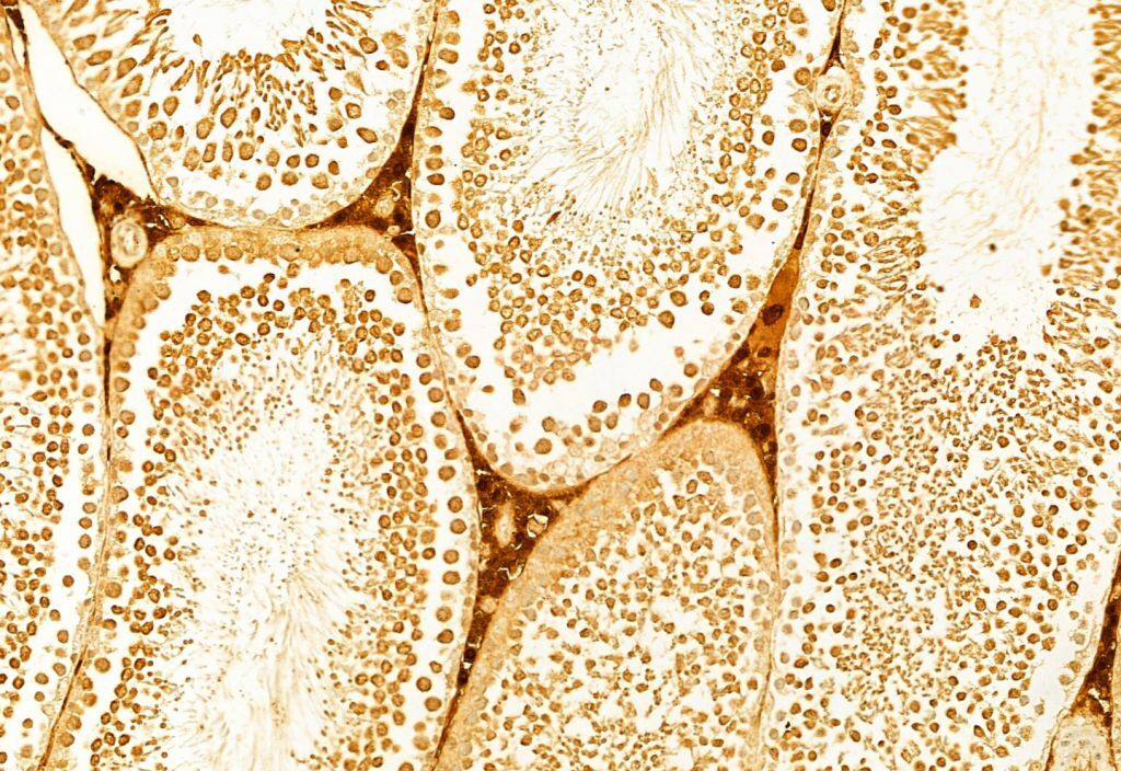
- Immunohistochemistry analysis of KEAP1 in rat testis tissue. The sample was formaldehyde fixed and a heat mediated antigen retrieval step in citrate buffer was performed. Samples were incubated with KEAP1 polyclonal antibody (Product # PA5-99434) using a dilution of 1:100 (4°C overnight) followed by HRP conjugated anti-Rabbit secondary antibody.

- Submitted by
- Invitrogen Antibodies (provider)
- Main image

- Experimental details
- Immunohistochemistry analysis of KEAP1 in mouse spleen tissue. The sample was formaldehyde fixed and a heat mediated antigen retrieval step in citrate buffer was performed. Samples were incubated with KEAP1 polyclonal antibody (Product # PA5-99434) using a dilution of 1:100 (4°C overnight) followed by HRP conjugated anti-Rabbit secondary antibody.
- Submitted by
- Invitrogen Antibodies (provider)
- Main image

- Experimental details
- Immunohistochemistry analysis of KEAP1 in mouse skin tissue. The sample was formaldehyde fixed and a heat mediated antigen retrieval step in citrate buffer was performed. Samples were incubated with KEAP1 polyclonal antibody (Product # PA5-99434) using a dilution of 1:100 (4°C overnight) followed by HRP conjugated anti-Rabbit secondary antibody.
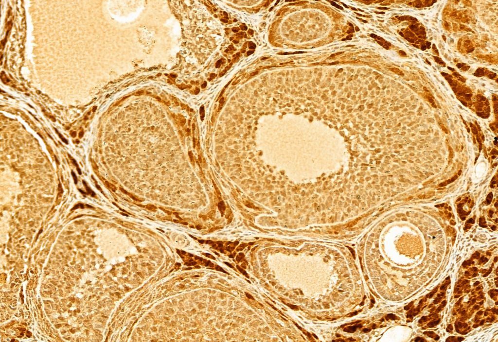
- Submitted by
- Invitrogen Antibodies (provider)
- Main image

- Experimental details
- Immunohistochemistry analysis of KEAP1 in rat ovary tissue. The sample was formaldehyde fixed and a heat mediated antigen retrieval step in citrate buffer was performed. Samples were incubated with KEAP1 polyclonal antibody (Product # PA5-99434) using a dilution of 1:100 (4°C overnight) followed by HRP conjugated anti-Rabbit secondary antibody.

- Submitted by
- Invitrogen Antibodies (provider)
- Main image
- Experimental details
- Immunohistochemistry analysis of KEAP1 in mouse liver tissue. The sample was formaldehyde fixed and a heat mediated antigen retrieval step in citrate buffer was performed. Samples were incubated with KEAP1 polyclonal antibody (Product # PA5-99434) using a dilution of 1:100 (4°C overnight) followed by HRP conjugated anti-Rabbit secondary antibody.

- Submitted by
- Invitrogen Antibodies (provider)
- Main image
- Experimental details
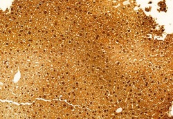
- Immunohistochemistry analysis of paraffin-embedded KEAP1 in human Heart muscle tissue sections. Antigen retrieval was performed using citrate buffer. Samples were blocked with blocking buffer (1.5 hr, 22°C), incubated with KEAP1 polyclonal antibody (Product # PA5-99434) using a dilution of 1:100 (1.5 hr, 22°C), followed by HRP conjugated goat anti-rabbit.
Supportive validation
- Submitted by
- Invitrogen Antibodies (provider)
- Main image
- Experimental details
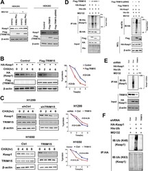
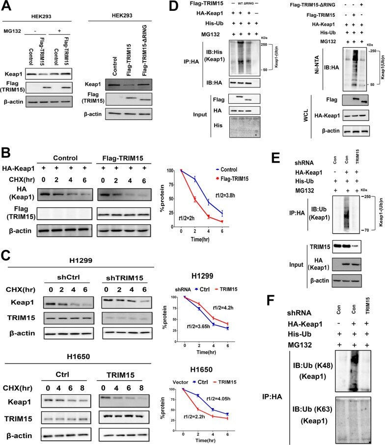
- Fig. 4 TRIM15 promotes Keap1 ubiquitination and degradation. A Immunoblot analysis of Keap1, Flag-TRIM15, and beta-actin in HEK293 cells transfected with expression vector for Flag-TRIM15 or with empty vector, with or without treatment of 10 muM MG132. Mutations of Flag-TRIM15-DeltaRING impaired the ability of TRIM15 to degrade Keap1 protein in HEK293 cells. B HEK293 cells were transfected with the indicated plasmids. The cells were treated with 50 mug/ml CHX for indicated time periods. Densitometry analysis performed on corresponding immunoblots to assess Keap1 half-life in the indicated conditions. C H1299 or H1650 cells were transfected with the indicated plasmids, treated with 50 mug/ml CHX, harvested at different time points, and then immunoblotted with using the indicated antibodies. Densitometry analysis performed on corresponding immunoblots to assess Keap1 half-life in the indicated conditions. D HEK293 cells were transfected with Flag-TRIM15, Flag-TRIM15-DeltaRING, HA-Keap1, and His-ubiquitin plasmids, and the cell lysates were subjected to immunoprecipitation using anti-HA antibodies (left panel) or Ni-NTA pull-down (right panel) under denaturing conditions, followed by immunoblotting with the indicated antibodies. Overexpression of wild-type but not the mutant TRIM15-DeltaRING promoted ubiquitination of Keap1. E Knockdown of endogenous TRIM15 decreased the ubiquitination of HA-Keap1 in HEK293 cells analyzed by in vitro ubiquitination assays. F Cell lysates prepare
- Submitted by
- Invitrogen Antibodies (provider)
- Main image
- Experimental details
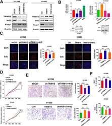
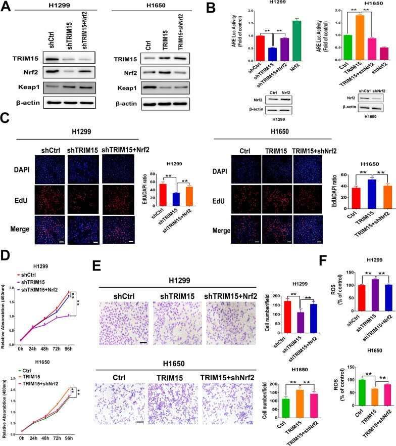
- Fig. 6 TRIM15-mediated Nrf2 signaling regulates growth and invasion in NSCLC cells in vitro. A Western blot analyses of TRIM15, Nrf2, Keap1, and Nrf2 target NQO1 in H1299 cells with TRIM15 knockdown with or without subsequent Nrf2 overexpression and H1650 cells overexpressing TRIM15 with or without subsequent knockdown of Nrf2. B ARE Luc reporter activity assessed in H1299 cells expressing shTRIM15, sh TRIM15 + Nrf2 or H1650 cells overexpressing TRIM15 with or without subsequent knockdown of Nrf2. Up-regulating of Nrf2 expression in H1299 cells or down-regulating of Nrf2 expression in H1650 cells was set as a control. C - F Cell proliferation ( C , D ), invasion ( E ) and ROS formation ( F ) in H1299 cells with or without shTRIM15 and Nrf2 rescue or in H1650 cells with or without TRIM15 overexpression and shNrf2 rescue. Statistical analyses were performed by two-tailed unpaired Student's t -test. ** P < 0.01
- Submitted by
- Invitrogen Antibodies (provider)
- Main image
- Experimental details
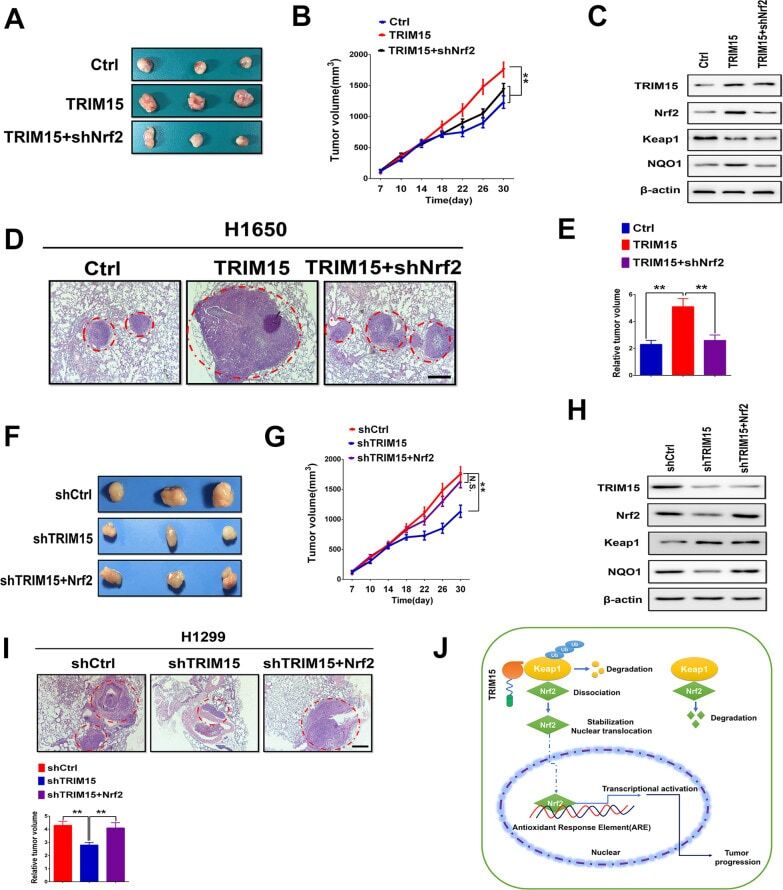
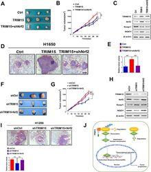
- Fig. 7 TRIM15 mediated increase in Nrf2 regulates growth and invasion in vivo. A Nude mice were randomized into three groups and subcutaneously injected with H1650 cells that had been transfected with control (empty vector), TRIM15, or TRIM15 + shNrf2 plasmids. Tumors formed in nude mice were collected 30 days after grafting, and the tumor weight were measured. B Measurement of tumor volume in experimental groups over time. C Western blotting analysis was performed to evaluate the levels of TRIM15, Nrf2, Keap1, and NQO1 in harvested tumors. D , E Up-regulation of TRIM15 significantly promoted lung metastasis in H1650 xenograft nude mice models, whereas the suppression of Nrf2 prevented the tumor metastasis of TRIM15 overexpressing cells. Representative pictures of the lung metastases in nude mice by H&E staining. Quantification of lung metastases in all groups. Scale bar: 200 mum. F , G A representative image of tumor growth in nude mice subcutaneously inoculated with H1299 cells tranfected with shCtrl, shTRIM15 or shTRIM15 + Nrf2 plasmids. Tumor volumes were measured on the indicated days. H Western blotting analysis was performed to evaluate the levels of TRIM15, Nrf2, Keap1, and NQO1 in xenograft tumors. I Representative pictures of the lung metastases in nude mice by H&E staining. Quantification of lung metastases in all groups. Scale bar: 200 mum. J TRIM15 was significantly upregulated in NSCLC and that increased TRIM15 was associated with poor survival. TRIM15 promoted