 Explore
 Validate
 Learn
Learn Western blot
 Other assay
Antibody data
- Antibody Data
- Antigen structure
- References [1]
- Comments [0]
- Validations
- Other assay [1]
Submit
Validation data
Reference
Comment
Report error
- Product number
- 720288 - Provider product page

- Provider
- Invitrogen Antibodies
- Product name
- BDKRB2 Polyclonal Antibody
- Antibody type
- Polyclonal
- Antigen
- Synthetic peptide
- Description
- These Polyclonal antibodies are of rabbit origin developed by immunizing animals with proteins or peptides. The polyclonal antibody is purified by affinity purification from the rabbit sera generated after immunizing the rabbits with a specific type of protein or peptide. The purified antibody is tested for its functionality in various relevant research applications. The antibody is developed for Research Use Only and is non-hazardous or non-infectious in nature. This antibody is predicted to react with Rat, Mouse, Pig and Cat.
- Reactivity
- Human, Mouse, Rat
- Host
- Rabbit
- Isotype
- IgG
- Vial size
- 100 μg
- Concentration
- 0.5 mg/mL
- Storage
- Store at 4°C short term. For long term storage, store at -20°C, avoiding freeze/thaw cycles.
Submitted references Heteromerization fingerprints between bradykinin B2 and thromboxane TP receptors in native cells.
Dagher OK, Jaffa MA, Habib A, Ziyadeh FN, Jaffa AA
PloS one 2019;14(5):e0216908
PloS one 2019;14(5):e0216908
No comments: Submit comment
Supportive validation
- Submitted by
- Invitrogen Antibodies (provider)
- Main image
- Experimental details
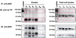
- Fig 5 B2R-TP interactions in RASMC as revealed by co-IP followed by SDS PAGE. RASMC lysates were immunoprecipitated with anti-B2R followed by immunoblotting with anti-TP (upper panels) and anti-B2R (lower panels) antibodies, successively. E and L represent eluates and matching lysates of unstimulated RASMC (E1, L1), or RASMC that were stimulated with BK 10-11 M (E2, L2), IBOP 10-7 M (E3, L3), or [BK 10-11 M + IBOP 10-7 M] (E4, L4) for 10 min. EM and LM represent eluates and matching lysates of mock co-IP condition. (EB) represents eluates of a control co-IP condition, whereby Dynabeads-protein-A-anti-B2R were incubated with PBS instead of RASMC lysates. Images are representative of three qualitatively similar independent experiments. A denatured broad molecular weight protein ladder was loaded in parallel (upper and lower left-hand lanes).
