 Explore
 Validate
 Learn
Learn Western blot
 Immunocytochemistry
Antibody data
- Antibody Data
- Antigen structure
- References [0]
- Comments [0]
- Validations
- Immunocytochemistry [3]
- Immunohistochemistry [1]
Submit
Validation data
Reference
Comment
Report error
- Product number
- 720275 - Provider product page

- Provider
- Invitrogen Antibodies
- Product name
- mGluR2 Polyclonal Antibody
- Antibody type
- Polyclonal
- Antigen
- Synthetic peptide
- Description
- These Polyclonal antibodies are of rabbit origin developed by immunizing animals with proteins or peptides. The polyclonal antibody is purified by affinity purification from the rabbit sera generated after immunizing the rabbits with a specific type of protein or peptide. The purified antibody is tested for its functionality in various relevant research applications. The antibody is developed for Research Use Only and is non-hazardous or non-infectious in nature. This antibody is predicted to react with Monkey, Rabbit and Bovine.
- Reactivity
- Human
- Host
- Rabbit
- Isotype
- IgG
- Vial size
- 100 µg
- Concentration
- 0.5 mg/mL
- Storage
- Store at 4°C short term. For long term storage, store at -20°C, avoiding freeze/thaw cycles.
No comments: Submit comment
Supportive validation
- Submitted by
- Invitrogen Antibodies (provider)
- Main image

- Experimental details
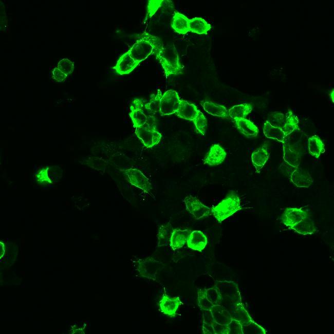
- HEK 293 cells were grown on poly-L-lysine-coated coverslips overnight. Cells were then transiently transfected with mGLUR2. After 24 h, cells were fixed with 4% paraformaldehyde and 0.2% picric acid in phosphate buffer (pH 6.9) for 30 min at room temperature and washed several times with phosphate buffer. Specimens were permeabilized with ice-cold methanol and then incubated with Anti-mGLUR2 Rabbit Polyclonal Antibody (Product # 720275 at a concentration of 2 µg/mL). Cells were then incubated with Alexa488-conjugated secondary antibody for 2 h at room temperature, mounted and examined using a laser scanning confocal microscope. Note, strong staining of transfected cells and no staining of adjacent non-transfected cells.
- Submitted by
- Invitrogen Antibodies (provider)
- Main image
- Experimental details
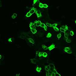
- HEK 293 cells were grown on poly-L-lysine-coated coverslips overnight. Cells were then transiently transfected with mGLUR2. After 24 h, cells were fixed with 4% paraformaldehyde and 0.2% picric acid in phosphate buffer (pH 6.9) for 30 min at room temperature and washed several times with phosphate buffer. Specimens were permeabilized with ice-cold methanol and then incubated with Anti-mGLUR2 Rabbit Polyclonal Antibody (Product # 720275 at a concentration of 2 µg/mL). Cells were then incubated with Alexa488-conjugated secondary antibody for 2 h at room temperature, mounted and examined using a laser scanning confocal microscope. Note, strong staining of transfected cells and no staining of adjacent non-transfected cells.

- Submitted by
- Invitrogen Antibodies (provider)
- Main image

- Experimental details
- HEK 293 cells were grown on poly-L-lysine-coated coverslips overnight. Cells were then transiently transfected with mGLUR2. After 24 h, cells were fixed with 4% paraformaldehyde and 0.2% picric acid in phosphate buffer (pH 6.9) for 30 min at room temperature and washed several times with phosphate buffer. Specimens were permeabilized with ice-cold methanol and then incubated with Anti-mGLUR2 Rabbit Polyclonal Antibody (Product # 720275 at a concentration of 2 µg/mL). Cells were then incubated with Alexa488-conjugated secondary antibody for 2 h at room temperature, mounted and examined using a laser scanning confocal microscope. Note, strong staining of transfected cells and no staining of adjacent non-transfected cells.

Supportive validation
- Submitted by
- Invitrogen Antibodies (provider)
- Main image
- Experimental details
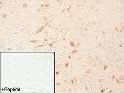
- Sections of human cerebral cortex were dewaxed, microwaved in citric acid and incubated with Anti-mGLUR2 Rabbit Polyclonal Antibody (Product # 720275 at a concentration of 2 µg/mL). Sections were then sequentially treated with biotinylated anti-rabbit IgG and AB solution. Sections were then developed in AEC and lightly counterstained with hematoxylin. Inset, for adsorption controls the antibody was incubated with 10 µg/mL of the peptide used for immunizations (+ Peptide). Note, staining of neuronal somata and dendrites is completely neutralized by preincubation of the antibody with its immunizing peptide.
