Explore
 Validate
 Learn
Learn Western blot
Antibody data
- Antibody Data
- Antigen structure
- References [0]
- Comments [0]
- Validations
- Western blot [1]
- Immunocytochemistry [2]
- Immunohistochemistry [1]
Submit
Validation data
Reference
Comment
Report error
- Product number
- 702592 - Provider product page

- Provider
- Invitrogen Antibodies
- Product name
- mGLuR2 Recombinant Rabbit Monoclonal Antibody (15H9L5)
- Antibody type
- Monoclonal
- Antigen
- Synthetic peptide
- Description
- This antibody is predicted to react with Monkey, Rabbit, Bovine. Recombinant rabbit monoclonal antibodies are produced using in vitro expression systems. The expression systems are developed by cloning in the specific antibody DNA sequences from immunoreactive rabbits. Then, individual clones are screened to select the best candidates for production. The advantages of using recombinant rabbit monoclonal antibodies include: better specificity and sensitivity, lot-to-lot consistency, animal origin-free formulations, and broader immunoreactivity to diverse targets due to larger rabbit immune repertoire.
- Reactivity
- Human, Rat
- Host
- Rabbit
- Isotype
- IgG
- Antibody clone number
- 15H9L5
- Vial size
- 100 μg
- Concentration
- 0.5 mg/mL
- Storage
- Store at 4°C short term. For long term storage, store at -20°C, avoiding freeze/thaw cycles.
No comments: Submit comment
Supportive validation
- Submitted by
- Invitrogen Antibodies (provider)
- Main image
- Experimental details
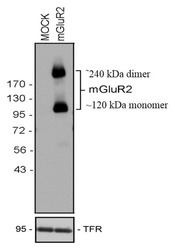
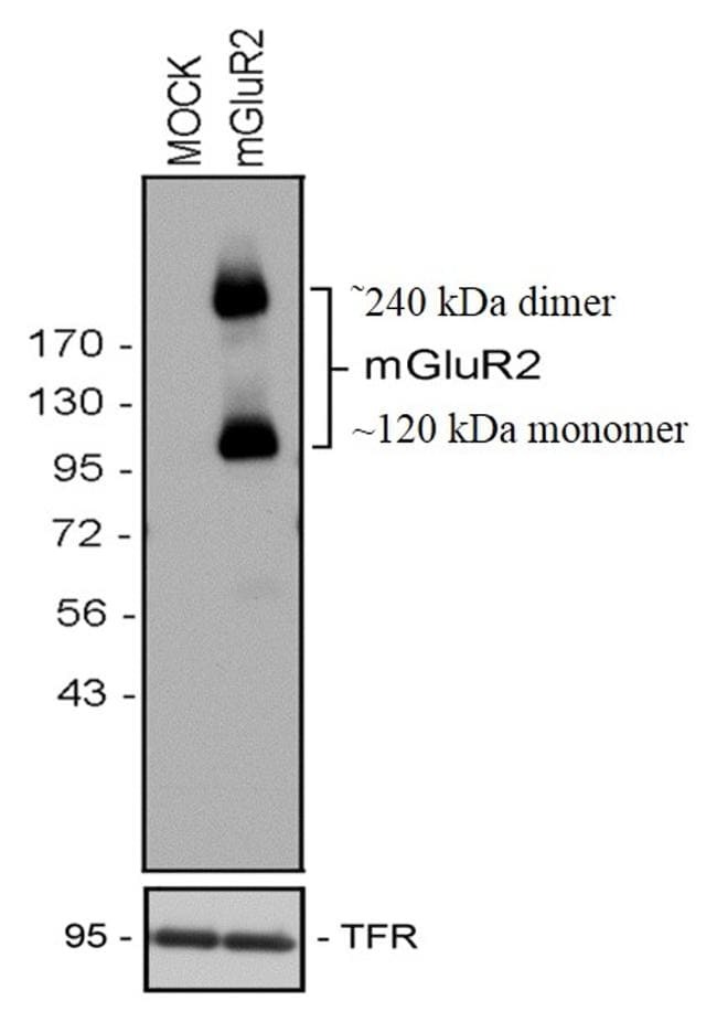
- Western blot analysis was performed on HEK-293 cells stably transfected with human mGLUR2 (Lane 2) and Mock (Lane 1). Receptors were eluted from the beads using SDS sample buffer for 20 min at 45 °C. Extracts were separated on 7.5% SDS-polyacrylamide gels and blotted onto PVDF membranes. The blot was probed with Anti-mGLuR2 Recombinant Rabbit Monoclonal Antibody (Product # 702592, 1 µg/mL, 1:500 dilution). A 120 kDa band and a 240 kDa band corresponding to the monomeric and dimeric forms of mGLuR2 was observed in the transfected cell line.
Supportive validation
- Submitted by
- Invitrogen Antibodies (provider)
- Main image

- Experimental details
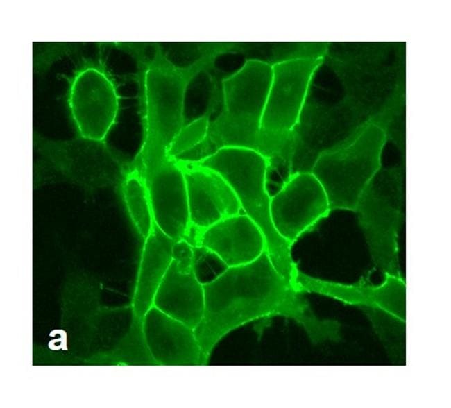
- For immunofluorescence analysis, HEK-293 cells were stably transfected with human mGLuR2. After 24hr of transfection the cells were fixed and permeabilized for detection of endogenous mGLuR2 using Anti-mGLuR2 Recombinant Rabbit Monoclonal Antibody (Product # 702592, 1 µg/mL, 1:500 dilution) and labeled with Goat anti-Rabbit IgG (H+L) Superclonal™ Secondary Antibody, Alexa Fluor® 488 conjugate. Panel a) shows representative cells that were stained for detection of membrane localization of mGLuR2 on the transfected cells. The images were captured at 40X magnification.

- Submitted by
- Invitrogen Antibodies (provider)
- Main image

- Experimental details
- For immunofluorescence analysis, HEK-293 cells were stably transfected with human mGLuR2. After 24hr of transfection the cells were fixed and permeabilized for detection of endogenous mGLuR2 using Anti-mGLuR2 Recombinant Rabbit Monoclonal Antibody (Product # 702592, 1 µg/mL, 1:500 dilution) and labeled with Goat anti-Rabbit IgG (H+L) Superclonal™ Secondary Antibody, Alexa Fluor® 488 conjugate. Panel a) shows representative cells that were stained for detection of membrane localization of mGLuR2 on the transfected cells. The images were captured at 40X magnification.
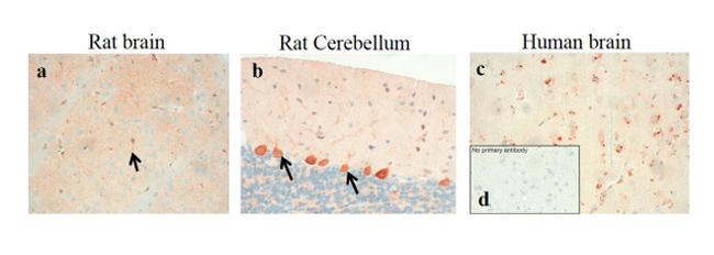
Supportive validation
- Submitted by
- Invitrogen Antibodies (provider)
- Main image
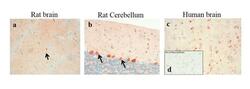
- Experimental details
- Sections of Rat Brain, Rat Cerebellum and Human Brain were dewaxed, microwaved in citric acid and incubated with Anti-mGLuR2 Recombinant Rabbit Monoclonal Antibody (Product # 702592, 1 µg/mL, 1:500 dilution). Sections were then sequentially treated with biotinylated Anti-rabbit IgG and AB solution. Sections were further developed in AEC and lightly counterstained with hematoxylin. Panel a, b & c) shows the specific localization of mGLuR2 in neuronal stomata and dendrites in Rat Brain, Rat Cerebellum and Human Brain respectively. Panel d) shows the no primary antibody control.