Explore
 Validate
 Learn
Learn Western blot
Antibody data
- Antibody Data
- Antigen structure
- References [0]
- Comments [0]
- Validations
- Western blot [2]
- Immunocytochemistry [1]
- Immunohistochemistry [1]
Submit
Validation data
Reference
Comment
Report error
- Product number
- AGC-011-200UL - Provider product page

- Provider
- Invitrogen Antibodies
- Product name
- mGluR2 (extracellular) Polyclonal Antibody
- Antibody type
- Polyclonal
- Antigen
- Other
- Reactivity
- Human, Mouse, Rat
- Host
- Rabbit
- Isotype
- IgG
- Vial size
- 200 µL
- Concentration
- 0.8 mg/mL
- Storage
- -20° C, Avoid Freeze/Thaw Cycles
No comments: Submit comment
Supportive validation
- Submitted by
- Invitrogen Antibodies (provider)
- Main image
- Experimental details
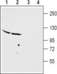
- Western blot analysisof rat cerebellum (lanes 1 and 3) and cortex (lanes 2 and 4) membranes: - 1,2. Anti-mGluR2 (extracellular) Antibody (#AGC-011), (1:400).3,4. Anti-mGluR2 (extracellular) Antibody , preincubated with mGluR2 (extracellular) Blocking Peptide (#BLP-GC011).

- Submitted by
- Invitrogen Antibodies (provider)
- Main image
- Experimental details
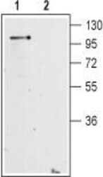
- Western blot analysisof mouse brainmembranes: - 1. Anti-mGluR2 (extracellular) Antibody (#AGC-011), (1:200). 2. Anti-mGluR2 (extracellular) Antibody , preincubated with mGluR2 (extracellular) Blocking Peptide (#BLP-GC011).

Supportive validation
- Submitted by
- Invitrogen Antibodies (provider)
- Main image
- Experimental details
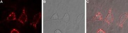
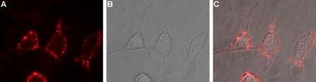
- Expression of mGluR2 in rat PC12 cells - Cell surface detection of mGluR2 in live intact rat PC12 pheochromocytoma cells. A. Extracellular staining of cells with Anti-mGluR2 (extracellular) Antibody (#AGC-011), (1:100), followed by goat Anti-rabbit-AlexaFluor-594 secondary Antibody (red). B. Live view of the cells. C. Merge of A and B.
Supportive validation
- Submitted by
- Invitrogen Antibodies (provider)
- Main image
- Experimental details
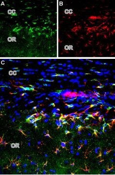
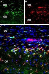
- Expression of mGluR2 in rat brain - Immunohistochemical staining of perfusion-fixed brain frozen sections using Anti-mGluR2 (extracellular) Antibody (#AGC-011). A. mGluR2 (green) is visualized in the corpus callosum (CC) and hippocampal stratum oriens (OR). B. Glial fibrillary acidic protein (GFAP) (red), a marker of astrocytes. C. Merge of the two images demonstrates expression of mGluR2 in astrocytes. DAPI is used as the nuclear counterstain (blue).