Explore
 Validate
 Learn
Learn Western blot
 Immunohistochemistry
Antibody data
- Antibody Data
- Antigen structure
- References [4]
- Comments [0]
- Validations
- Western blot [3]
- Immunoprecipitation [1]
Submit
Validation data
Reference
Comment
Report error
- Product number
- NB100-2305 - Provider product page

- Provider
- Novus Biologicals
- Proper citation
- Novus Cat#NB100-2305, RRID:AB_2136662
- Product name
- Rabbit Polyclonal EPLIN Antibody
- Antibody type
- Polyclonal
- Description
- Immunogen affinity purified.
- Reactivity
- Human
- Host
- Rabbit
- Isotype
- IgG
- Vial size
- 100 ul
- Concentration
- 1.0 mg/ml
- Storage
- Store at 4C. Do not freeze.
Submitted references Epidermal growth factor promotes protein degradation of epithelial protein lost in neoplasm (EPLIN), a putative metastasis suppressor, during epithelial-mesenchymal transition.
Quantitative protein and mRNA profiling shows selective post-transcriptional control of protein expression by vasopressin in kidney cells.
EPLIN downregulation promotes epithelial-mesenchymal transition in prostate cancer cells and correlates with clinical lymph node metastasis.
Extracellular signal-regulated kinase/mitogen-activated protein kinase regulates actin organization and cell motility by phosphorylating the actin cross-linking protein EPLIN.
Zhang S, Wang X, Iqbal S, Wang Y, Osunkoya AO, Chen Z, Chen Z, Shin DM, Yuan H, Wang YA, Zhau HE, Chung LW, Ritenour C, Kucuk O, Wu D
The Journal of biological chemistry 2013 Jan 18;288(3):1469-79
The Journal of biological chemistry 2013 Jan 18;288(3):1469-79
Quantitative protein and mRNA profiling shows selective post-transcriptional control of protein expression by vasopressin in kidney cells.
Khositseth S, Pisitkun T, Slentz DH, Wang G, Hoffert JD, Knepper MA, Yu MJ
Molecular & cellular proteomics : MCP 2011 Jan;10(1):M110.004036
Molecular & cellular proteomics : MCP 2011 Jan;10(1):M110.004036
EPLIN downregulation promotes epithelial-mesenchymal transition in prostate cancer cells and correlates with clinical lymph node metastasis.
Zhang S, Wang X, Osunkoya AO, Iqbal S, Wang Y, Chen Z, Müller S, Chen Z, Josson S, Coleman IM, Nelson PS, Wang YA, Wang R, Shin DM, Marshall FF, Kucuk O, Chung LW, Zhau HE, Wu D
Oncogene 2011 Dec 15;30(50):4941-52
Oncogene 2011 Dec 15;30(50):4941-52
Extracellular signal-regulated kinase/mitogen-activated protein kinase regulates actin organization and cell motility by phosphorylating the actin cross-linking protein EPLIN.
Han MY, Kosako H, Watanabe T, Hattori S
Molecular and cellular biology 2007 Dec;27(23):8190-204
Molecular and cellular biology 2007 Dec;27(23):8190-204
No comments: Submit comment
Supportive validation
- Submitted by
- Novus Biologicals (provider)
- Main image

- Experimental details
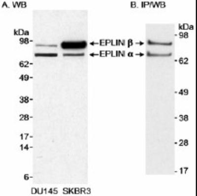
- Western Blot: EPLIN Antibody [NB100-2305] - Detection of Human EPLIN by Western Blot. Samples: A) Whole cell lysate (50 mcg) from DU145 and SKBR3 cells. B) EPLIN that was immunoprecipitated from 500 mcg of whole cell lysate from DU145 cells. Antibodies: Affinity purified rabbit anti-EPLIN antibody NB100-2035 was used at 0.3 mcg/ml (A) or 1 mcg/ml (B) for WB. In B, EPLIN was immunoprecipitated using another rabbit anti-EPLIN antibody. Detection: Chemiluminescence with exposure times of 30 minutes (A) or 30 seconds (B).
- Submitted by
- Novus Biologicals (provider)
- Main image
- Experimental details
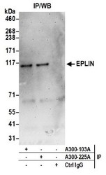
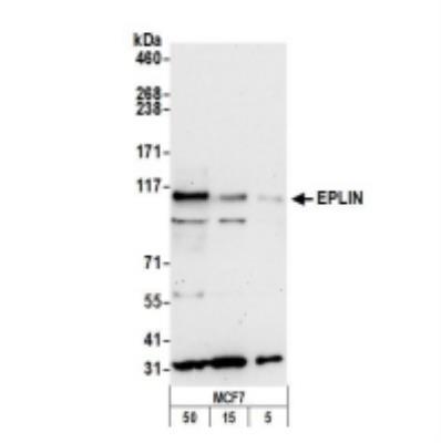
- Western Blot: EPLIN Antibody [NB100-2305] - Samples: Whole cell lysate (0.5 or 1.0 mg per IP reaction; 20% of IP loaded) from MCF7 cells prepared using NETN lysis buffer. Antibodies: Affinity purified rabbit anti-EPLIN antibody (lot was used at 1 ug/ml. Detection: Chemiluminescence with an exposure time of 3 minutes.
- Submitted by
- Novus Biologicals (provider)
- Main image
- Experimental details
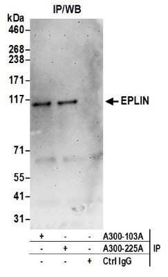
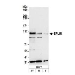
- Western Blot: EPLIN Antibody [NB100-2305] - Whole cell lysate from MCF7 (5, 15 and 50 ug) cells prepared using NETN lysis buffer. Antibody: Affinity purified rabbit anti-EPLIN antibody used for WB at 0.4 ug/ml. Detection: Chemiluminescence with an exposure time of 30 seconds.
Supportive validation
- Submitted by
- Novus Biologicals (provider)
- Main image
- Experimental details
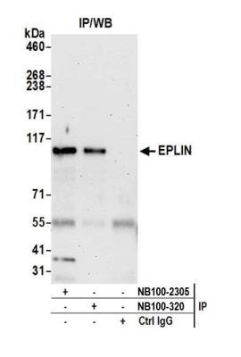
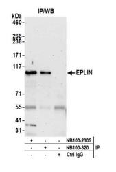
- Immunoprecipitation: EPLIN Antibody [NB100-2305] - Detection of human EPLIN by western blot of immunoprecipitates. Samples: Whole cell lysate (0.5 or 1.0 mg per IP reaction; 20% of IP loaded) from MCF-7 cells prepared using NETN lysis buffer. Antibodies: Affinity purified rabbit anti-EPLIN antibody NB100-2305 (lot NB100-2305-4) used for IP at 6 ug per reaction. EPLIN was also immunoprecipitated by rabbit anti-EPLIN antibody NB100-320. For blotting immunoprecipitated EPLIN, NB100-2305 was used at 1 ug/ml. Detection: Chemiluminescence with an exposure time of 75 seconds.