 Explore
 Validate
 Learn
Learn Western blot
Antibody data
- Antibody Data
- Antigen structure
- References [1]
- Comments [0]
- Validations
- Western blot [6]
Submit
Validation data
Reference
Comment
Report error
- Product number
- PA1-24435 - Provider product page

- Provider
- Invitrogen Antibodies
- Product name
- Anti-TFAM Polyclonal Antibody
- Antibody type
- Polyclonal
- Antigen
- Synthetic peptide
- Description
- Recommended positive controls: bEND.3, Hep3B, HepG2, Jurkat, K562. Predicted reactivity: Cow: 92%; Dog: 83%; Guinea Pig: 75%; Horse: 92%; Mouse: 75%; Pig: 92%; Rabbit: 83%. By Western blot, PA1-24435 detects the ~ 29 kDa TFAM protein. Store product as a concentrated solution. Centrifuge briefly prior to opening the vial.
- Reactivity
- Human, Mouse, Rat
- Host
- Rabbit
- Isotype
- IgG
- Vial size
- 50 µg
- Concentration
- 1 mg/mL
- Storage
- Store at 4°C short term. For long term storage, store at -20°C, avoiding freeze/thaw cycles.
Submitted references Endurance exercise as a countermeasure for aging.
Lanza IR, Short DK, Short KR, Raghavakaimal S, Basu R, Joyner MJ, McConnell JP, Nair KS
Diabetes 2008 Nov;57(11):2933-42
Diabetes 2008 Nov;57(11):2933-42
No comments: Submit comment
Supportive validation
- Submitted by
- Invitrogen Antibodies (provider)
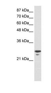
- Main image

- Experimental details
- Western blot analysis of TFAM using a TFAM polyclonal antibody (Product # PA1-24435).

- Submitted by
- Invitrogen Antibodies (provider)
- Main image

- Experimental details
- Western blot analysis of TFAM in bEND3 cell lysate at 10 µg (lane 1) and 20 µg (lane 2) using a TFAM polyclonal antibody (Product # PA1-24435).

- Submitted by
- Invitrogen Antibodies (provider)
- Main image

- Experimental details
- Western blot analysis of TFAM in Jurkat cell lysate using a TFAM polyclonal antibody (Product # PA1-24435).
- Submitted by
- Invitrogen Antibodies (provider)
- Main image

- Experimental details
- Western Blot analysis of TFAM was performed by loading Jurkat cells. Proteins were transferred to a membrane and probed with a TFAM Polyclonal Antibody (Product # PA1-24435) at a dilution of 0.2-1 µg/mL.

- Submitted by
- Invitrogen Antibodies (provider)
- Main image

- Experimental details
- Knockdown of mtTFA was achieved by transfecting HeLa cells with mtTFA specific siRNAs (Silencer® select Product # s14000, s14001). Western blot analysis (Fig. a) was performed membrane enriched extracts from the HeLa knockdown cells (lane 3), non-specific scrambled siRNA transfected cells (lane 2) and untransfected cells (lane 1). The blot was probed with mtTFA Polyclonal Antibody (Product # PA1-24435, 1 µg/mL) and Goat anti-Rabbit IgG (H+L) Superclonal™ Recombinant Secondary Antibody, HRP (Product # A27036, 1:4000 dilution). Densitometric analysis of this western blot is shown in histogram (Fig. b). Decrease in signal upon siRNA mediated knock down confirms that antibody is specific to mtTFA.

- Submitted by
- Invitrogen Antibodies (provider)
- Main image

- Experimental details
- Western blot was performed using Anti-mtTFA Rabbit Polyclonal Antibody (Product # PA1-24435) and a 26 kDa band corresponding to mtTFA was observed in cell lines and tissues tested. Membrane enriched extracts (30 µg lysate) of HeLa (Lane 1), T-47D (Lane 2), MDA-MB-231 (Lane 3), Caco-2 (Lane 4), Mouse Heart (Lane 5) and Rat Heart (Lane 6) were electrophoresed using NuPAGE™ 12% Bis-Tris Protein Gel (Product # NP0342BOX). Resolved proteins were then transferred onto a nitrocellulose membrane (Product # IB23001) by iBlot® 2 Dry Blotting System (Product # IB21001). The blot was probed with the primary antibody (1 µg/mL) and detected by chemiluminescence with Goat anti-Rabbit IgG (H+L) Superclonal™ Recombinant Secondary Antibody, HRP (Product # A27036, 1:4000 dilution) using the iBright FL 1000 (Product # A32752). Chemiluminescent detection was performed using Novex® ECL Chemiluminescent Substrate Reagent Kit (Product # WP20005).
